Thu, Oct 1, 2026
Volume 11 - Continuous Publishing
Iran J Neurosurg 2025, 11 - Continuous Publishing: 0-0 |
Back to browse issues page



Download citation:
BibTeX | RIS | EndNote | Medlars | ProCite | Reference Manager | RefWorks
Send citation to:


BibTeX | RIS | EndNote | Medlars | ProCite | Reference Manager | RefWorks
Send citation to:
Fahim F, Mehmandoost M, Abbasi H, Sharifzadeh S, Bahri A M, Oveisi S. Middle Meningeal Artery Embolization in Septated Chronic Subdural Hematoma: A Narrative Review. Iran J Neurosurg 2025; 11 : 24
URL: http://irjns.org/article-1-495-en.html
URL: http://irjns.org/article-1-495-en.html
Farzan Fahim *1 

 , Mahdi Mehmandoost2 , Hesan Abbasi2 , Shahrzad Sharifzadeh1 , Amir Mohammad Bahri1 , Sayeh Oveisi1
, Mahdi Mehmandoost2 , Hesan Abbasi2 , Shahrzad Sharifzadeh1 , Amir Mohammad Bahri1 , Sayeh Oveisi1
1- Functional Neurosurgery Research Center, Research Institute of Functional Neurosurgery, Shohada Tajrish Comprehensive Neurosurgical Center of Excellence, Shahid Beheshti University of Medical Sciences, Tehran, Iran.
2- Student Research Committee, School of Medicine, Shahid Beheshti University of Medical Sciences, Tehran, Iran.
2- Student Research Committee, School of Medicine, Shahid Beheshti University of Medical Sciences, Tehran, Iran.
Keywords: Chronic subdural hematoma, Septated cSDH, Middle meningeal artery embolization (MMAE), Recurrence, Neovascularization, Minimally invasive therapy
Full Text [PDF 1000 kb]
(310 Downloads)
| Abstract (HTML) (1500 Views)
Full Text: (159 Views)
1. Introduction
Chronic subdural hematoma (cSDH) refers to the accumulation of blood between the arachnoid and dura mater, typically developing over a period of three weeks or longer [1]. It is more prevalent in the elderly population [2], with an estimated incidence ranging from 1.72 to 20.6 cases per 100,000 persons per year—a figure of considerable concern given the associated morbidities and mortalities [2]. A distinct subtype of cSDH, known as septated cSDH, is characterized by the organization of subdural blood clots into fibrinous membranes, which exhibit neovascularization and increased vascular permeability [3]. This neovascularization contributes to the formation of fragile vessels that can exacerbate hematoma expansion, leading to recurrent episodes [4], thereby complicating treatment strategies. Management approaches for cSDH include conservative treatment, craniotomy, burr hole evacuation, and craniostomy [5].
Middle meningeal artery embolization (MMAE) is a minimally invasive procedure aimed at occluding the vascular supply to pathological dural membranes, thereby reducing the risk of recurrent hemorrhage in cases of cSDH. While MMAE has emerged as a promising alternative to conventional surgical interventions, its therapeutic efficacy in septated cSDH a subtype associated with a higher recurrence rate—remains uncertain and warrants further investigation [6, 7]. This review seeks to provide a comprehensive analysis of the existing literature on septated cSDH and the potential role of MMAE as a treatment modality.
2. Methods and Materials/pateints
We conducted a targeted narrative review focusing on MMAE for septated chronic subdural hematoma (sCSDH). A comprehensive literature search was performed in PubMed, Scopus, and Web of Science from January 2000 to July 2025. Additional searches were done in Google Scholar and by hand-screening reference lists of relevant articles. The search combined Medical Subject Headings (MeSH) and free-text terms:
(“chronic subdural hematoma” OR cSDH) AND (septated OR membranous OR multiloculated OR organized) AND (“middle meningeal artery” OR MMA) AND (embolization OR “endovascular therapy” OR neurointervention). Only peer-reviewed human studies were included. Two authors independently screened titles/abstracts, then full-texts. Disagreements were resolved by consensus.
Eligibility criteria
Inclusion criteria: Human studies on cSDH with focus on septated/membranous/multiloculated subtypes. MMAE as primary or adjunct to burr-hole, craniotomy, or twist-drill evacuation.
Outcomes including recurrence, hematoma resolution (radiologic/clinical), complications, and technical aspects (access route, embolic agent). Study types: RCTs, cohort, case-control, large case series, systematic/narrative reviews with relevant mechanistic insight (2000–2025). Exclusion criteria: Non-septated cSDH without subgroup analysis. Abstract-only reports, animal/in-vitro studies.
Pathophysiology of septated CSDH
The defining pathological feature of septated cSDH (sCSDH) is the formation of fibrin septations and thickened inner membranes, which result from chronic inflammatory processes and recurrent microhemorrhages within the subdural space [8]. These septa alter the fluid dynamics of the hematoma, preventing uniform liquefaction and forming physical barriers that obstruct complete drainage during surgical intervention [8].
A cSDH is characterized as septated based on computed tomography (CT) imaging criteria, specifically when one or more linear hyperdense structures were observed within the predominantly hypodense hematoma [7]. Imaging characteristics of sCSDH in MRI of some cases are internal septations, mixed signal intensity, dural thickening, diffuse inflammatory enhancement, and absence of abnormal enhancing mass [9].
3. Current Treatment Modalities for cSDH
Surgical interventions
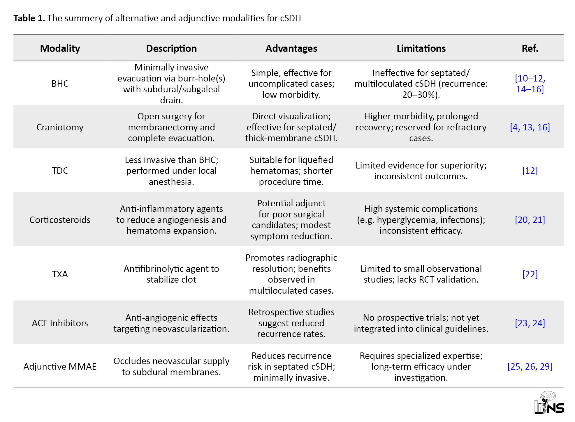
Surgical evacuation remains the primary treatment modality for symptomatic cSDH, particularly in patients presenting with significant mass effect or neurological deficits. Among the available techniques, burr-hole craniostomy (BHC) is the most widely performed procedure due to its simplicity, minimal invasiveness, and generally favorable outcomes in uncomplicated cases [10]. The procedure involves creating one or more burr-holes to facilitate hematoma drainage, typically followed by the placement of a subdural or subgaleal drain to minimize recurrence risk [11].
For select patients especially those with liquefied hematomas or undergoing the procedure under local anesthesia twist-drill craniostomy (TDC) may provide a less invasive alternative with comparable efficacy. However, evidence regarding its superiority or equivalence remains inconclusive [12]. Conversely, craniotomy, a more invasive approach, is reserved for cases where thick outer membranes, septations, or solid hematoma components impede adequate drainage via burr-hole techniques [13]. Despite its higher risk profile, craniotomy enables direct visualization, membranectomy, and complete evacuation in refractory cases.
Traditional surgical techniques, however, often demonstrate reduced effectiveness in managing septated or multiloculated cSDH, where the hematoma is compartmentalized by fibrous membranes and trabeculations. These septations hinder uniform drainage, frequently resulting in incomplete evacuation and predisposing patients to persistent collections and recurrence. Studies indicate recurrence rates of 20% to 30% in septated cases, even following technically sufficient surgical intervention [14, 15]. Notably, Nakaguchi et al. identified the presence of internal membranes as a significant predictor of postoperative recurrence [16].
Limitations in septated CSDH
Septated cSDH poses a distinct clinical and surgical challenge due to its unique pathophysiological and structural characteristics. The fibrous septa, composed of proliferative membranes rich in inflammatory and angiogenic markers, compartmentalize the hematoma cavity and hinder uniform decompression [17]. These internal partitions frequently trap residual hematoma fluid or clotted blood, resulting in persistent mass effect and ongoing symptoms, even after apparent surgical evacuation.
Furthermore, the inability to fully collapse or remove these compartments through burr-hole techniques alone often necessitates repeat surgery or conversion to craniotomy, thereby increasing patient morbidity and prolonging hospitalization [18]. These challenges are particularly pronounced in elderly or medically fragile patients, who may have limited tolerance for multiple interventions or general anesthesia. As a result, septated cSDH has increasingly been recognized as a refractory form of the condition, often resistant to conventional monotherapeutic strategies [19].
Pharmacologic and adjuvant therapies
Given the limitations of surgical monotherapy, various adjuvant medical therapies have been investigated to improve treatment efficacy and reduce recurrence risk. Among these, corticosteroids have garnered significant attention for their potential role in modulating hematoma stability. Their anti-inflammatory properties may theoretically suppress the expression of proangiogenic cytokines and inhibit neomembrane formation, thereby reducing hematoma expansion [20]. However, clinical trials have yielded inconsistent results. In a randomized controlled trial (RCT), Hutchinson et al. (2020) reported a modest reduction in the need for surgical intervention with dexamethasone; however, this benefit was accompanied by an increased incidence of systemic complications, including hyperglycemia and infections [21]. Consequently, corticosteroids are now considered a potentially beneficial adjunct in select patients, particularly those who are poor surgical candidates, rather than a primary treatment strategy.
Tranexamic acid (TXA), an antifibrinolytic agent, has emerged as a promising pharmacological option due to its ability to inhibit fibrinolysis within the hematoma, thereby promoting clot stability. Kageyama et al. reported favorable radiographic resolution and symptom improvement in patients treated with TXA, including those with multiloculated hematomas [22]. However, current evidence remains limited to observational studies and small case series, highlighting the need for further randomized trials to validate its efficacy and safety.
Another class of pharmacological agents gaining attention is angiotensin-converting enzyme inhibitors (ACE-Is). Given their antiangiogenic and anti-inflammatory effects, ACE-Is may theoretically reduce neovascular permeability and decrease recurrence rates in cSDH [23]. Retrospective cohort studies have suggested a potential correlation between ACE-I use and lower recurrence rates; however, prospective validation is required before ACE-Is can be integrated into routine clinical management [24].
Need for alternative and adjunctive modalities
Given the persistent recurrence, surgical challenges, and limited pharmacologic success in managing septated cSDH, there is increasing recognition of the need for alternative or adjunctive treatment strategies that address its underlying pathophysiological mechanisms namely, chronic inflammation, fragile neovasculature, and progressive hematoma expansion. MMAE has emerged as a particularly promising approach in this context. By selectively occluding the vascular supply to the outer neomembrane, MMAE seeks to disrupt the inflammatory-angiogenic cycle that perpetuates hematoma persistence and recurrence [25].
As discussed in subsequent sections, MMAE may serve as either a standalone therapy particularly in recurrent or medically inoperable cases or as an adjunct to surgical evacuation in septated hematomas. Preliminary evidence suggests that integrating MMAE with surgical intervention may enhance outcomes by preventing neomembrane revascularization and mitigating the risk of blood reaccumulation in residual compartments (Table 1) [26].
4. MMAE: Mechanism and Technique
Middle meningeal artery (MMA) embolization aims to devascularize the subdural membranes of chronic subdural hematomas (cSDHs), effectively shifting the balance from persistent blood leakage and accumulation toward enhanced reabsorption [27]. MMAE can control hemorrhage originating from the cSDH membrane and, in some cases, facilitate spontaneous hematoma resolution without direct drainage. However, symptomatic hematomas may still necessitate surgical removal This technique has emerged as a promising therapeutic option for cSDH, either as a standalone procedure or as an adjunct to surgical intervention. The efficacy of MMAE in cSDH has been explored in multiple RCTs [28]. Given the rich vascularity of hematoma membranes, MMAE has been proposed as a potentially beneficial approach for patients with septated cSDH [28].
Although surgical evacuation remains the standard treatment for cSDH, recurrence rates remain substantial, occurring in approximately 10–20% of cases following a single evacuation [29]. Over the past few years, the number of studies reporting MMAE efficacy has increased significantly. The three primary indications for standalone MMAE include failure of conservative management in asymptomatic or mildly symptomatic patients, advanced age or concurrent use of antiplatelet/anticoagulant therapy (as MMAE is less invasive than surgery), and prevention of recurrence in patients with prior disease relapse following surgical evacuation [29]. Notably, septated cSDH presents additional challenges, as patients with septations exhibit lower rates of complete evacuation and a higher likelihood of recurrence. Reports indicate that MMAE has significantly reduced recurrence rates in septated cases compared to non-septated cases (SEP vs no-SEP: 3.1% vs 23.4%, P=0.017) [7]. Septation formation is thought to occur during the later stages of cSDH and may represent a more mature and densely neovascularized environment factors that make MMAE particularly suited for treatment [7].
Most MMAE procedures are performed via transfemoral access (TFA), though this approach can pose challenges due to the risk of access site complications and the requirement for postoperative bed rest [30]. In contrast, transradial access (TRA), which is commonly employed in cardiac catheterization, is gaining popularity in neurointerventional procedures due to its lower risk of site complications and reduced postoperative bed rest requirements [30].
Various particulate embolic agents are utilized in MMAE, functioning as flow-directed materials delivered via catheter to induce either temporary or permanent arterial occlusion. These agents may be therapeutically effective in managing cSDH [31]. Among them, polyvinyl alcohol (PVA) and Embosphere particles promote occlusion albeit with a potential risk of recanalization by adhering to vessel walls within the neurovasculature and inducing a chronic inflammatory response that ultimately leads to fibrosis. In contrast, gelatin sponge provides a temporary occlusive effect via mechanical obstruction, reducing intravascular flow and serving as a structural scaffold for thrombus formation [31]. Among embolic materials, PVA particles are the most frequently used embolic agents in studies evaluating MMAE for cSDH treatment [31]. For elderly patients, liquid embolic agents can be infused into the distal vasculature through a wedged catheter position and injected under pressure to create a cast of the subdural membranes, often resulting in reflux into adjacent meningeal branches (Table 2) [27].
Evidence for MMAE in septated cSDH
cSDH is a complex condition with an incidence rate ranging from 1.7 to 20.6 per 100,000 individuals [32]. Given the clinical significance of this disease, we reviewed multiple studies evaluating the efficacy of MMAE in septated cSDH.
In one case, an 86-year-old man receiving warfarin for ventricular fibrillation was diagnosed with cSDH via CT. Despite undergoing bilateral burr-hole drainage, complete evacuation was not achieved. Two months later, the patient was hospitalized due to fever and refractory headache, indicating recurrence of cSDH. Following craniotomy, MMAE was performed, leading to complete hematoma resolution within six months [33].
Another case involved a 5-year-old female with idiopathic cardiomyopathy who underwent heart transplantation. Unfortunately, left ventricular thrombosis resulted in ischemic stroke, with subsequent CT imaging revealing an enlarging cSDH. MMAE was performed at seven months, facilitating resolution of the hematoma [34].
A case series analysis further examined three patients who underwent initial burr-hole irrigation or drainage. All three subsequently received MMAE at different time intervals, demonstrating a reduction in hematoma size following embolization [35].
In a study by Link et al. MMAE was performed in six patients with cSDH, leading to a significant reduction in lesion size from 25 mm at baseline to 6 mm after 24 weeks. Additionally, another investigation involving 80 patients (median age: 68 years) reported septated membrane cSDH in 35% of cases. Of these, 45% underwent burr-hole evacuation, 18.8% received craniotomy, and the remainder received MMAE. The findings highlighted that MMAE significantly reduced hematoma width and recurrence risk [7].
Furthermore, a separate study evaluated 28 patients with cSDH treated with surgery alone or surgery plus MMAE. Results indicated that MMAE effectively prevented recurrence in these cases [28]. Another study assessed MMAE in patients with a median age of 75 years, showing an 87% reduction in lesion size without mortality [36].
Another retrospective study categorized cSDH patients into membranous (61%) and non-membranous (73.7%) groups. Those treated with MMAE and burr-hole evacuation exhibited a 10% recurrence rate [37].
Organized subdural hematoma (OSDH), a rare variant of cSDH, was successfully treated with craniotomy followed by MMAE. A 61-year-old patient underwent twist-drill craniotomy, yet hematoma recurrence necessitated embolization, ultimately leading to resolution [9].
Additionally, treatment selection considerations were highlighted in another study. While craniotomy remains an effective approach, it carries inherent risks of bleeding and recurrence. In two reported cases, a 71-year-old man and a 77-year-old man underwent burr-hole irrigation, MMAE, and craniotomy sequentially, both achievzing complete neurological recovery without recurrence [38].
Kadono et al. further supported MMAE’s efficacy in cSDH management, assessing two cases. In the first, an 83-year-old man with left cSDH drainage was hospitalized due to dysarthria and dysplasia. Initial craniotomy was followed by MMAE, resulting in complete hematoma resolution after six months. Similarly, another 83-year-old patient with cSDH underwent burr-hole perforation followed by MMAE, leading to symptom resolution within three months (Table 3) [39].
5. Conclusion
MMAE represents a transformative advancement in managing septated cSDH, a subtype characterized by fibrin septations and high recurrence rates following conventional surgical interventions. Current evidence demonstrates that adjunctive or standalone MMAE significantly reduces recurrence rates in septated cSDH (3.1% vs 23.4% in non-embolized cases), primarily by disrupting the angiogenic-inflammatory cycle driving hematoma persistence. Technical refinements, such as TRA and optimized embolic agents (e.g., PVA, gelatin sponge), enhance procedural safety, particularly in elderly or high-risk populations. Despite these benefits, challenges persist, including variability in embolic protocols, the necessity for neurointerventional expertise, and limited long-term outcome data. Emerging studies position MMAE as a paradigm-shifting adjunct to surgery, offering improved outcomes in refractory or recurrent cases. However, prospective randomized trials are imperative to establish standardized protocols, validate long-term efficacy, and solidify MMAE’s role in clinical guidelines. This review underscores MMAE as a pivotal strategy in the evolving management of septated cSDH, bridging gaps left by traditional therapies while advocating for multidisciplinary collaboration to refine its application.
Ethical Considerations
Compliance with ethical guidelines
Ethics approval, consent to participate, and consent to publish are not applicable to this manuscript, as it is a narrative review of existing literature and does not involve human/animal subjects or original data collection.
Funding
This research received no specific grant from funding agencies in the public, commercial, or not-for-profit sectors.
Authors' contributions
Conception and design: Farzan Fahim, Mohammad Ali Nazari and Mahdi Mehmandoost; Data Collection: Hesan Abbasi; Drafting the article: Mohammad Ali Nazari, Mahdi Mehmandoost, Sayeh Oveisi and Amirmohammad Bahri; Critically revising the article: Farzan Fahim; Reviewing submitted version of manuscript: Farzan Fahim; Final Approl: All authors.
Conflict of interest
The authors declared no conflict of interest.
References
Chronic subdural hematoma (cSDH) refers to the accumulation of blood between the arachnoid and dura mater, typically developing over a period of three weeks or longer [1]. It is more prevalent in the elderly population [2], with an estimated incidence ranging from 1.72 to 20.6 cases per 100,000 persons per year—a figure of considerable concern given the associated morbidities and mortalities [2]. A distinct subtype of cSDH, known as septated cSDH, is characterized by the organization of subdural blood clots into fibrinous membranes, which exhibit neovascularization and increased vascular permeability [3]. This neovascularization contributes to the formation of fragile vessels that can exacerbate hematoma expansion, leading to recurrent episodes [4], thereby complicating treatment strategies. Management approaches for cSDH include conservative treatment, craniotomy, burr hole evacuation, and craniostomy [5].
Middle meningeal artery embolization (MMAE) is a minimally invasive procedure aimed at occluding the vascular supply to pathological dural membranes, thereby reducing the risk of recurrent hemorrhage in cases of cSDH. While MMAE has emerged as a promising alternative to conventional surgical interventions, its therapeutic efficacy in septated cSDH a subtype associated with a higher recurrence rate—remains uncertain and warrants further investigation [6, 7]. This review seeks to provide a comprehensive analysis of the existing literature on septated cSDH and the potential role of MMAE as a treatment modality.
2. Methods and Materials/pateints
We conducted a targeted narrative review focusing on MMAE for septated chronic subdural hematoma (sCSDH). A comprehensive literature search was performed in PubMed, Scopus, and Web of Science from January 2000 to July 2025. Additional searches were done in Google Scholar and by hand-screening reference lists of relevant articles. The search combined Medical Subject Headings (MeSH) and free-text terms:
(“chronic subdural hematoma” OR cSDH) AND (septated OR membranous OR multiloculated OR organized) AND (“middle meningeal artery” OR MMA) AND (embolization OR “endovascular therapy” OR neurointervention). Only peer-reviewed human studies were included. Two authors independently screened titles/abstracts, then full-texts. Disagreements were resolved by consensus.
Eligibility criteria
Inclusion criteria: Human studies on cSDH with focus on septated/membranous/multiloculated subtypes. MMAE as primary or adjunct to burr-hole, craniotomy, or twist-drill evacuation.
Outcomes including recurrence, hematoma resolution (radiologic/clinical), complications, and technical aspects (access route, embolic agent). Study types: RCTs, cohort, case-control, large case series, systematic/narrative reviews with relevant mechanistic insight (2000–2025). Exclusion criteria: Non-septated cSDH without subgroup analysis. Abstract-only reports, animal/in-vitro studies.
Pathophysiology of septated CSDH
The defining pathological feature of septated cSDH (sCSDH) is the formation of fibrin septations and thickened inner membranes, which result from chronic inflammatory processes and recurrent microhemorrhages within the subdural space [8]. These septa alter the fluid dynamics of the hematoma, preventing uniform liquefaction and forming physical barriers that obstruct complete drainage during surgical intervention [8].
A cSDH is characterized as septated based on computed tomography (CT) imaging criteria, specifically when one or more linear hyperdense structures were observed within the predominantly hypodense hematoma [7]. Imaging characteristics of sCSDH in MRI of some cases are internal septations, mixed signal intensity, dural thickening, diffuse inflammatory enhancement, and absence of abnormal enhancing mass [9].
3. Current Treatment Modalities for cSDH
Surgical interventions
Surgical evacuation remains the primary treatment modality for symptomatic cSDH, particularly in patients presenting with significant mass effect or neurological deficits. Among the available techniques, burr-hole craniostomy (BHC) is the most widely performed procedure due to its simplicity, minimal invasiveness, and generally favorable outcomes in uncomplicated cases [10]. The procedure involves creating one or more burr-holes to facilitate hematoma drainage, typically followed by the placement of a subdural or subgaleal drain to minimize recurrence risk [11].
For select patients especially those with liquefied hematomas or undergoing the procedure under local anesthesia twist-drill craniostomy (TDC) may provide a less invasive alternative with comparable efficacy. However, evidence regarding its superiority or equivalence remains inconclusive [12]. Conversely, craniotomy, a more invasive approach, is reserved for cases where thick outer membranes, septations, or solid hematoma components impede adequate drainage via burr-hole techniques [13]. Despite its higher risk profile, craniotomy enables direct visualization, membranectomy, and complete evacuation in refractory cases.
Traditional surgical techniques, however, often demonstrate reduced effectiveness in managing septated or multiloculated cSDH, where the hematoma is compartmentalized by fibrous membranes and trabeculations. These septations hinder uniform drainage, frequently resulting in incomplete evacuation and predisposing patients to persistent collections and recurrence. Studies indicate recurrence rates of 20% to 30% in septated cases, even following technically sufficient surgical intervention [14, 15]. Notably, Nakaguchi et al. identified the presence of internal membranes as a significant predictor of postoperative recurrence [16].
Limitations in septated CSDH
Septated cSDH poses a distinct clinical and surgical challenge due to its unique pathophysiological and structural characteristics. The fibrous septa, composed of proliferative membranes rich in inflammatory and angiogenic markers, compartmentalize the hematoma cavity and hinder uniform decompression [17]. These internal partitions frequently trap residual hematoma fluid or clotted blood, resulting in persistent mass effect and ongoing symptoms, even after apparent surgical evacuation.
Furthermore, the inability to fully collapse or remove these compartments through burr-hole techniques alone often necessitates repeat surgery or conversion to craniotomy, thereby increasing patient morbidity and prolonging hospitalization [18]. These challenges are particularly pronounced in elderly or medically fragile patients, who may have limited tolerance for multiple interventions or general anesthesia. As a result, septated cSDH has increasingly been recognized as a refractory form of the condition, often resistant to conventional monotherapeutic strategies [19].
Pharmacologic and adjuvant therapies
Given the limitations of surgical monotherapy, various adjuvant medical therapies have been investigated to improve treatment efficacy and reduce recurrence risk. Among these, corticosteroids have garnered significant attention for their potential role in modulating hematoma stability. Their anti-inflammatory properties may theoretically suppress the expression of proangiogenic cytokines and inhibit neomembrane formation, thereby reducing hematoma expansion [20]. However, clinical trials have yielded inconsistent results. In a randomized controlled trial (RCT), Hutchinson et al. (2020) reported a modest reduction in the need for surgical intervention with dexamethasone; however, this benefit was accompanied by an increased incidence of systemic complications, including hyperglycemia and infections [21]. Consequently, corticosteroids are now considered a potentially beneficial adjunct in select patients, particularly those who are poor surgical candidates, rather than a primary treatment strategy.
Tranexamic acid (TXA), an antifibrinolytic agent, has emerged as a promising pharmacological option due to its ability to inhibit fibrinolysis within the hematoma, thereby promoting clot stability. Kageyama et al. reported favorable radiographic resolution and symptom improvement in patients treated with TXA, including those with multiloculated hematomas [22]. However, current evidence remains limited to observational studies and small case series, highlighting the need for further randomized trials to validate its efficacy and safety.
Another class of pharmacological agents gaining attention is angiotensin-converting enzyme inhibitors (ACE-Is). Given their antiangiogenic and anti-inflammatory effects, ACE-Is may theoretically reduce neovascular permeability and decrease recurrence rates in cSDH [23]. Retrospective cohort studies have suggested a potential correlation between ACE-I use and lower recurrence rates; however, prospective validation is required before ACE-Is can be integrated into routine clinical management [24].
Need for alternative and adjunctive modalities
Given the persistent recurrence, surgical challenges, and limited pharmacologic success in managing septated cSDH, there is increasing recognition of the need for alternative or adjunctive treatment strategies that address its underlying pathophysiological mechanisms namely, chronic inflammation, fragile neovasculature, and progressive hematoma expansion. MMAE has emerged as a particularly promising approach in this context. By selectively occluding the vascular supply to the outer neomembrane, MMAE seeks to disrupt the inflammatory-angiogenic cycle that perpetuates hematoma persistence and recurrence [25].
As discussed in subsequent sections, MMAE may serve as either a standalone therapy particularly in recurrent or medically inoperable cases or as an adjunct to surgical evacuation in septated hematomas. Preliminary evidence suggests that integrating MMAE with surgical intervention may enhance outcomes by preventing neomembrane revascularization and mitigating the risk of blood reaccumulation in residual compartments (Table 1) [26].
4. MMAE: Mechanism and Technique
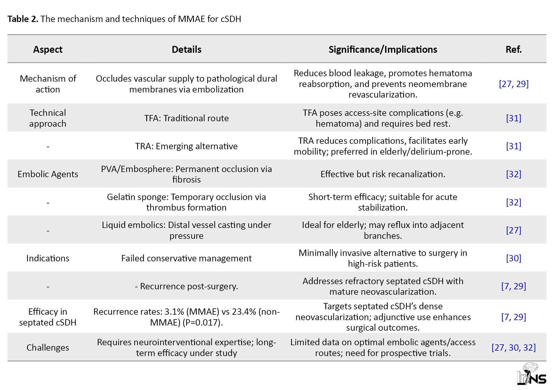
Middle meningeal artery (MMA) embolization aims to devascularize the subdural membranes of chronic subdural hematomas (cSDHs), effectively shifting the balance from persistent blood leakage and accumulation toward enhanced reabsorption [27]. MMAE can control hemorrhage originating from the cSDH membrane and, in some cases, facilitate spontaneous hematoma resolution without direct drainage. However, symptomatic hematomas may still necessitate surgical removal This technique has emerged as a promising therapeutic option for cSDH, either as a standalone procedure or as an adjunct to surgical intervention. The efficacy of MMAE in cSDH has been explored in multiple RCTs [28]. Given the rich vascularity of hematoma membranes, MMAE has been proposed as a potentially beneficial approach for patients with septated cSDH [28].
Although surgical evacuation remains the standard treatment for cSDH, recurrence rates remain substantial, occurring in approximately 10–20% of cases following a single evacuation [29]. Over the past few years, the number of studies reporting MMAE efficacy has increased significantly. The three primary indications for standalone MMAE include failure of conservative management in asymptomatic or mildly symptomatic patients, advanced age or concurrent use of antiplatelet/anticoagulant therapy (as MMAE is less invasive than surgery), and prevention of recurrence in patients with prior disease relapse following surgical evacuation [29]. Notably, septated cSDH presents additional challenges, as patients with septations exhibit lower rates of complete evacuation and a higher likelihood of recurrence. Reports indicate that MMAE has significantly reduced recurrence rates in septated cases compared to non-septated cases (SEP vs no-SEP: 3.1% vs 23.4%, P=0.017) [7]. Septation formation is thought to occur during the later stages of cSDH and may represent a more mature and densely neovascularized environment factors that make MMAE particularly suited for treatment [7].
Most MMAE procedures are performed via transfemoral access (TFA), though this approach can pose challenges due to the risk of access site complications and the requirement for postoperative bed rest [30]. In contrast, transradial access (TRA), which is commonly employed in cardiac catheterization, is gaining popularity in neurointerventional procedures due to its lower risk of site complications and reduced postoperative bed rest requirements [30].
Various particulate embolic agents are utilized in MMAE, functioning as flow-directed materials delivered via catheter to induce either temporary or permanent arterial occlusion. These agents may be therapeutically effective in managing cSDH [31]. Among them, polyvinyl alcohol (PVA) and Embosphere particles promote occlusion albeit with a potential risk of recanalization by adhering to vessel walls within the neurovasculature and inducing a chronic inflammatory response that ultimately leads to fibrosis. In contrast, gelatin sponge provides a temporary occlusive effect via mechanical obstruction, reducing intravascular flow and serving as a structural scaffold for thrombus formation [31]. Among embolic materials, PVA particles are the most frequently used embolic agents in studies evaluating MMAE for cSDH treatment [31]. For elderly patients, liquid embolic agents can be infused into the distal vasculature through a wedged catheter position and injected under pressure to create a cast of the subdural membranes, often resulting in reflux into adjacent meningeal branches (Table 2) [27].
Evidence for MMAE in septated cSDH
cSDH is a complex condition with an incidence rate ranging from 1.7 to 20.6 per 100,000 individuals [32]. Given the clinical significance of this disease, we reviewed multiple studies evaluating the efficacy of MMAE in septated cSDH.
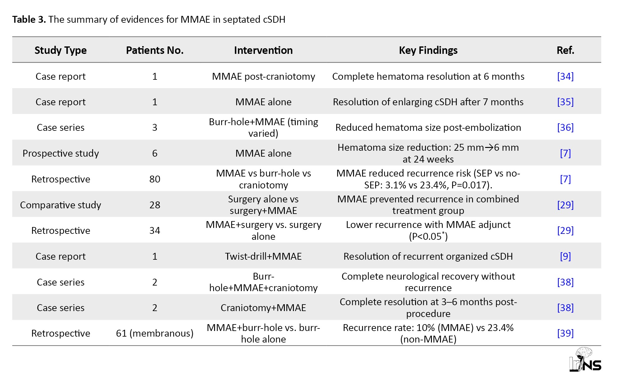
In one case, an 86-year-old man receiving warfarin for ventricular fibrillation was diagnosed with cSDH via CT. Despite undergoing bilateral burr-hole drainage, complete evacuation was not achieved. Two months later, the patient was hospitalized due to fever and refractory headache, indicating recurrence of cSDH. Following craniotomy, MMAE was performed, leading to complete hematoma resolution within six months [33].
Another case involved a 5-year-old female with idiopathic cardiomyopathy who underwent heart transplantation. Unfortunately, left ventricular thrombosis resulted in ischemic stroke, with subsequent CT imaging revealing an enlarging cSDH. MMAE was performed at seven months, facilitating resolution of the hematoma [34].
A case series analysis further examined three patients who underwent initial burr-hole irrigation or drainage. All three subsequently received MMAE at different time intervals, demonstrating a reduction in hematoma size following embolization [35].
In a study by Link et al. MMAE was performed in six patients with cSDH, leading to a significant reduction in lesion size from 25 mm at baseline to 6 mm after 24 weeks. Additionally, another investigation involving 80 patients (median age: 68 years) reported septated membrane cSDH in 35% of cases. Of these, 45% underwent burr-hole evacuation, 18.8% received craniotomy, and the remainder received MMAE. The findings highlighted that MMAE significantly reduced hematoma width and recurrence risk [7].
Furthermore, a separate study evaluated 28 patients with cSDH treated with surgery alone or surgery plus MMAE. Results indicated that MMAE effectively prevented recurrence in these cases [28]. Another study assessed MMAE in patients with a median age of 75 years, showing an 87% reduction in lesion size without mortality [36].
Another retrospective study categorized cSDH patients into membranous (61%) and non-membranous (73.7%) groups. Those treated with MMAE and burr-hole evacuation exhibited a 10% recurrence rate [37].
Organized subdural hematoma (OSDH), a rare variant of cSDH, was successfully treated with craniotomy followed by MMAE. A 61-year-old patient underwent twist-drill craniotomy, yet hematoma recurrence necessitated embolization, ultimately leading to resolution [9].
Additionally, treatment selection considerations were highlighted in another study. While craniotomy remains an effective approach, it carries inherent risks of bleeding and recurrence. In two reported cases, a 71-year-old man and a 77-year-old man underwent burr-hole irrigation, MMAE, and craniotomy sequentially, both achievzing complete neurological recovery without recurrence [38].
Kadono et al. further supported MMAE’s efficacy in cSDH management, assessing two cases. In the first, an 83-year-old man with left cSDH drainage was hospitalized due to dysarthria and dysplasia. Initial craniotomy was followed by MMAE, resulting in complete hematoma resolution after six months. Similarly, another 83-year-old patient with cSDH underwent burr-hole perforation followed by MMAE, leading to symptom resolution within three months (Table 3) [39].
5. Conclusion
MMAE represents a transformative advancement in managing septated cSDH, a subtype characterized by fibrin septations and high recurrence rates following conventional surgical interventions. Current evidence demonstrates that adjunctive or standalone MMAE significantly reduces recurrence rates in septated cSDH (3.1% vs 23.4% in non-embolized cases), primarily by disrupting the angiogenic-inflammatory cycle driving hematoma persistence. Technical refinements, such as TRA and optimized embolic agents (e.g., PVA, gelatin sponge), enhance procedural safety, particularly in elderly or high-risk populations. Despite these benefits, challenges persist, including variability in embolic protocols, the necessity for neurointerventional expertise, and limited long-term outcome data. Emerging studies position MMAE as a paradigm-shifting adjunct to surgery, offering improved outcomes in refractory or recurrent cases. However, prospective randomized trials are imperative to establish standardized protocols, validate long-term efficacy, and solidify MMAE’s role in clinical guidelines. This review underscores MMAE as a pivotal strategy in the evolving management of septated cSDH, bridging gaps left by traditional therapies while advocating for multidisciplinary collaboration to refine its application.
Ethical Considerations
Compliance with ethical guidelines
Ethics approval, consent to participate, and consent to publish are not applicable to this manuscript, as it is a narrative review of existing literature and does not involve human/animal subjects or original data collection.
Funding
This research received no specific grant from funding agencies in the public, commercial, or not-for-profit sectors.
Authors' contributions
Conception and design: Farzan Fahim, Mohammad Ali Nazari and Mahdi Mehmandoost; Data Collection: Hesan Abbasi; Drafting the article: Mohammad Ali Nazari, Mahdi Mehmandoost, Sayeh Oveisi and Amirmohammad Bahri; Critically revising the article: Farzan Fahim; Reviewing submitted version of manuscript: Farzan Fahim; Final Approl: All authors.
Conflict of interest
The authors declared no conflict of interest.
References
- Hamou HA, Clusmann H, Schulz JB, Wiesmann M, Altiok E, Höllig A. Chronic subdural hematoma: Antithrombotics and thrombotic compli89hes Ärzteblatt International. 2022; 119(12):208. [DOI:10.3238/arztebl.m2022.0144] [PMID]
- Rauhala M, Helén P, Huhtala H, Heikkilä P, Iverson GL, Niskakangas T, et al. Chronic subdural hematoma-incidence, complications, and financial impact. Acta Neurochirurgica. 2020; 162(9):2033-43. [DOI:10.1007/s00701-020-04398-3] [PMID] [PMCID]
- Singh H, Patir R, Vaishya S, Miglani R, Gupta A, Kaur A. Endoscopic evacuation of septated chronic subdural hemorrhage-Technical considerations, results, and outcome. Surgical Neurology International. 2022; 13:8. [DOI:10.25259/SNI_963_2021] [PMID] [PMCID]
- Abecassis IJ, Kim LJ. Craniotomy for treatment of chronic subdural hematoma. Neurosurgery Clinics of North America. 2017; 28(2):229-37. [DOI:10.1016/j.nec.2016.11.005] [PMID]
- Deng J, Wang F, Wang H, Zhao M, Chen G, Shangguan H, et al. Efficacy of neuroendoscopic treatment for septated chronic subdural hematoma. Frontiers in Neurology. 2021; 12:765109. [DOI:10.3389/fneur.2021.765109] [PMID] [PMCID]
- Davies JM, Knopman J, Mokin M, Hassan AE, Harbaugh RE, Khalessi A, et al. Adjunctive middle meningeal artery embolization for subdural hematoma. The New England Journal of Medicine. 2024; 391(20):1890-900. [DOI:10.1056/NEJMoa2313472] [PMID] [PMCID]
- Martinez-Gutierrez JC, D’Amato SA, Zeineddine HA, Nahhas MI, Kole MJ, Kim HW, et al. Middle meningeal artery embolization of septated chronic subdural hematomas. Interventional Neuroradiology. 2023; 31(6):745-50. [DOI:10.1177/15910199231184521] [PMID] [PMCID]
- An Y, Cheng H, Wang X, Men Y, Yu J, Zhang G, et al. Comparative analysis of endoscopic-assisted burr hole craniostomy and two burr hole craniostomy in the treatment of septated chronic subdural hematoma. Frontiers in Neurology. 2025; 16:1540877. [DOI:10.3389/fneur.2025.1540877] [PMID] [PMCID]
- Xuan HD, Minh TP, Van HD, Thanh DL, Tuan DP, Quang LT. Preoperative middle meningeal artery embolization in the treatment of organized chronic subdural hematoma. Radiology Case Reports. 2024; 19(8):3569-73. [DOI:10.1016/j.radcr.2024.04.090] [PMID] [PMCID]
- Santarius T, Kirkpatrick PJ, Ganesan D, Chia HL, Jalloh I, Smielewski P, et al. Use of drains versus no drains after burr-hole evacuation of chronic subdural haematoma: A randomised controlled trial. Lancet. 2009; 374(9695):1067-73. [DOI:10.1016/S0140-6736(09)61115-6] [PMID]
- Weigel R, Schmiedek P, Krauss JK. Outcome of contemporary surgery for chronic subdural haematoma: Evidence based review. J Neurol Neurosurg Psychiatry. 2003;74(7):937-43. [DOI:10.1136/jnnp.74.7.937] [PMID] [PMCID]
- Lee KS. How to treat chronic subdural hematoma? Past and now. Journal of Neurology, Neurosurgery, and Psychiatry. 2019; 62(2):144-52. [DOI:10.3340/jkns.2018.0156] [PMID] [PMCID]
- Ivamoto HS, Lemos HP, Jr., Atallah AN. Surgical treatments for chronic subdural hematomas: A comprehensive systematic review. World Neurosurgery. 2016; 86:399-418. [DOI:10.1016/j.wneu.2015.10.025] [PMID]
- Lee JY, Ebel H, Ernestus RI, Klug N. Various surgical treatments of chronic subdural hematoma and outcome in 172 patients: Is membranectomy necessary? Surgical Neurology. 2004; 61(6):523-7. [DOI:10.1016/j.surneu.2003.10.026] [PMID]
- Shapey J, Glancz LJ, Brennan PM. Chronic subdural haematoma in the elderly: Is it time for a new paradigm in management? Current Geriatrics Reports. 2016; 5:71-7. [DOI:10.1007/s13670-016-0166-9] [PMID] [PMCID]
- Nakaguchi H, Tanishima T, Yoshimasu N. Factors in the natural history of chronic subdural hematomas that influence their postoperative recurrence. Journal of Neurosurgery. 2001; 95(2):256-62. [DOI:10.3171/jns.2001.95.2.0256] [PMID]
- Edlmann E, Giorgi-Coll S, Whitfield PC, Carpenter KLH, Hutchinson PJ. Pathophysiology of chronic subdural haematoma: inflammation, angiogenesis and implications for pharmacotherapy. Journal of Neuroinflammation. 2017; 14(1):108. [DOI:10.1186/s12974-017-0881-y] [PMID] [PMCID]
- de Araújo Silva DO, Matis GK, Costa LF, Kitamura MA, de Carvalho Junior EV, de Moura Silva M, et al. Chronic subdural hematomas and the elderly: Surgical results from a series of 125 cases: Old “horses” are not to be shot! Surgical Neurology International. 2012; 3:150. [DOI:10.4103/2152-7806.104744] [PMID] [PMCID]
- Brennan PM, Kolias AG, Joannides AJ, Shapey J, Marcus HJ, Gregson BA, et al. The management and outcome for patients with chronic subdural hematoma: a prospective, multicenter, observational cohort study in the United Kingdom. J Neurosurg. 2017;127(4):732-9. [DOI:10.3171/2016.8.JNS16134] [PMID]
- Miah IP, Herklots M, Roks G, Peul WC, Walchenbach R, Dammers R, et al. Dexamethasone therapy in symptomatic chronic subdural hematoma (DECSA-R): A retrospective evaluation of initial corticosteroid therapy versus primary surgery. Journal of Neurosurgery. 2020; 37(2):366-72. [DOI:10.1089/neu.2019.6541] [PMID]
- Hutchinson PJ, Edlmann E, Bulters D, Zolnourian A, Holton P, Suttner N, et al. Trial of dexamethasone for chronic subdural hematoma. The New England Journal of Medicine. 2020; 383(27):2616-27. [DOI:10.1056/NEJMoa2020473] [PMID]
- Kageyama H, Toyooka T, Tsuzuki N, Oka K. Nonsurgical treatment of chronic subdural hematoma with tranexamic acid. Journal of Neurosurgery. 2013; 119(2):332-7. [DOI:10.3171/2013.3.JNS122162] [PMID]
- Ishii Y, Sakamoto T, Ito R, Yanaga K. Anti-angiogenic therapy on hepatocellular carcinoma development and progression. The Journal of Surgical Research. 2010; 158(1):69-76. [DOI:10.1016/j.jss.2008.09.015] [PMID]
- Weigel R, Hohenstein A, Schlickum L, Weiss C, Schilling L. Angiotensin converting enzyme inhibition for arterial hypertension reduces the risk of recurrence in patients with chronic subdural hematoma possibly by an antiangiogenic mechanism. Neurosurgery. 2007; 61(4):788-92. [DOI:10.1227/01.NEU.0000298907.56012.E8] [PMID]
- Link TW, Boddu S, Paine SM, Kamel H, Knopman J. Middle meningeal artery embolization for chronic subdural hematoma: A series of 60 cases. Neurosurgery. 2019;85(6):801-7. [DOI:10.1093/neuros/nyy521] [PMID]
- Ng S, Derraz I, Boetto J, Dargazanli C, Poulen G, Gascou G, et al. Middle meningeal artery embolization as an adjuvant treatment to surgery for symptomatic chronic subdural hematoma: A pilot study assessing hematoma volume resorption. Journal of Neurointerventional Surgery. 2020; 12(7):695-9. [DOI:10.1136/neurintsurg-2019-015421] [PMID]
- Fiorella D, Arthur AS. Middle meningeal artery embolization for the management of chronic subdural hematoma. Journal of Neurointerventional Surgery. 2019; 11(9):912-5. [DOI:10.1136/neurintsurg-2019-014730] [PMID]
- Martinez-Gutierrez JC, Dawes BH, Zeineddine HA, Wroe WW, D’Amato SA, Kim HW, et al. Middle meningeal artery embolization reduces recurrence following surgery for septated chronic subdural hematomas. Clinical Neurology and Neurosurgery. 2024;240:108252. [DOI:10.1016/j.clineuro.2024.108252] [PMID]
- Omura Y, Ishiguro T. Middle meningeal artery embolization for chronic subdural hematoma: A systematic review.Frontiers in Neurology. 2023; 14:1259647. [DOI:10.3389/fneur.2023.1259647] [PMID] [PMCID]
- Yamamoto M, Fujiwara G, Takezawa H, Uzura Y, Yokoya S, Oka H. Transradial versus transfemoral access for middle meningeal artery embolization: Choice of the access route considering delirium in the elderly. Surgical Neurology International. 2024; 15:276. [DOI:10.25259/SNI_168_2024] [PMID] [PMCID]
- Tudor T, Capone S, Vivanco-Suarez J, Salem MM, Sioutas GS, Tonetti DA, et al. Middle meningeal artery embolization for chronic subdural hematoma: A review of established and emerging embolic agents. Stroke. 2023; 4(1):e000906. [DOI:10.1161/SVIN.123.000906] [PMID] [PMCID]
- Feghali J, Yang W, Huang J. Updates in chronic subdural hematoma: epidemiology, etiology, pathogenesis, treatment, and outcome. World Neurosurgery. 2020; 141:339-45. [DOI:10.1016/j.wneu.2020.06.140] [PMID]
- Sato M, Mochizuki Y, Fukuchi M, Fujii K. Middle meningeal artery embolization before craniotomy for infected organizing chronic subdural hematoma: A case report and review of the literature. Surgical Neurology International. 2022; 13:186. [DOI:10.25259/SNI_73_2022] [PMID] [PMCID]
- Shigematsu T, Dier E, Yaeger KA, Morgenstern PF, Glass LD, Kellner CP, et al. Middle meningeal artery embolization of a pediatric patient with progressive chronic subdural hematoma. Operative Neurosurgery. 2021; 21(3):E274-E7. [DOI:10.1093/ons/opab144] [PMID]
- Hashimoto T, Ohashi T, Watanabe D, Koyama S, Namatame H, Izawa H, et al. Usefulness of embolization of the middle meningeal artery for refractory chronic subdural hematomas. Surgical Neurology International. 2013; 4:104. [DOI:10.4103/2152-7806.116679] [PMID] [PMCID]
- Mureb MC, Kondziolka D, Shapiro M, Raz E, Nossek E, Haynes J, et al. DynaCT enhancement of subdural membranes after middle meningeal artery embolization: Insights into pathophysiology. World Neurosurgery. 2020; 139:e265-70. [DOI:10.1016/j.wneu.2020.03.188] [PMID]
- Weinberg JH, Akhter A, Zakeri A, Tanweer O, Zyck S, Tjoumakaris S, et al. Middle meningeal artery embolization for membranous versus nonmembranous subdural hematomas: A retrospective and multicenter cohort study. World Neurosurgery. 2023; 177:e680-e5. [DOI:10.1016/j.wneu.2023.06.122] [PMID]
- Yokoya S, Nishii S, Takezawa H, Katsumori T, Takagi Y, Goto Y, et al. Organized chronic subdural hematoma treated with middle meningeal artery embolization and small craniotomy: Two case reports. Asian Journal of Neurosurgery. 2020; 15(2):421-4. [DOI:10.4103/ajns.AJNS_341_19] [PMID] [PMCID]
- Kadono Y, Kajikawa R, Tsuzuki T, Kishima H. Middle meningeal artery embolization for organized chronic subdural hematoma combined with minimal evacuation surgery: Two case reports. Radiology Case Reports. 2024; 19(12):6328-32. [DOI:10.1016/j.radcr.2024.09.062] [PMID] [PMCID]
Type of Study: Review |
Subject:
Endovascular Neurosurgery
| Rights and Permissions | |
 |
This work is licensed under a Creative Commons Attribution-NonCommercial 4.0 International License. |

Copyright © The Author(s);
This is an open access article distributed under the terms of the Creative Commons Attribution License (CC-By-NC), which permits use, distribution, and reproduction in any medium, provided the original work is properly cited and is not used for commercial purposes.
Contact Information
This is an open access article distributed under the terms of the Creative Commons Attribution License (CC-By-NC), which permits use, distribution, and reproduction in any medium, provided the original work is properly cited and is not used for commercial purposes.
Contact Information
