Mon, Sep 28, 2026
Volume 11 - Continuous Publishing
Iran J Neurosurg 2025, 11 - Continuous Publishing: 0-0 |
Back to browse issues page



Download citation:
BibTeX | RIS | EndNote | Medlars | ProCite | Reference Manager | RefWorks
Send citation to:


BibTeX | RIS | EndNote | Medlars | ProCite | Reference Manager | RefWorks
Send citation to:
Köksal V, Demirel C, Van M, Tomakin F, Engin R. A Rare Case of Adult Thoracic SCIWORA: Complete Cord Transection Without Vertebral Injury. Iran J Neurosurg 2025; 11 : 21
URL: http://irjns.org/article-1-496-en.html
URL: http://irjns.org/article-1-496-en.html


1- Department of Neurosurgery, Faculty of Medicine, Samsun Training and Research Hospital, Samsun University, Samsun, Türkey.
Keywords: Spinal cord injury, SCIWORA, Thoracic spine, Adult trauma, Spinal cord transection, Magnetic resonance imaging
Full Text [PDF 2086 kb]
(279 Downloads)
| Abstract (HTML) (1192 Views)
Full Text: (135 Views)
1. Background and Importance
Spinal cord injury without radiographic abnormality (SCIWORA) was first introduced by Pang and Wilberger in 1982 to describe spinal cord injuries in pediatric patients whose radiographs and computed tomography (CT) scans showed no signs of vertebral fracture or dislocation [1]. The term has historically been associated with the pediatric population, particularly in relation to the cervical spine, due to the greater elasticity and flexibility of the immature spinal column [2].
With the advent of magnetic resonance imaging (MRI), our ability to detect intramedullary spinal cord damage has significantly improved, leading to the identification of SCIWORA-like presentations in adult patients as well [3]. However, in contrast to pediatric cases, adult cases of SCIWORA are rare and often debated due to the relative rigidity of the mature spinal column and reduced ligamentous laxity [4, 5].
The thoracic spine, being the most rigid and biomechanically stable segment of the vertebral column, is especially resistant to injury without associated osseous or ligamentous disruption. Consequently, thoracic SCIWORA in adults is exceedingly uncommon, with only a limited number of reported cases in the literature [6–9].
Herein, we present a rare case of an adult patient who developed complete paraplegia following a high-energy motor vehicle accident, in the absence of any thoracic vertebral fracture or dislocation. MRI subsequently revealed a full-thickness transection of the spinal cord at the T7–T8 level. This case underscores the importance of maintaining a high index of suspicion for spinal cord injury in trauma patients with neurological deficits but unremarkable initial radiographs or CT scans. It also contributes to the limited body of literature documenting thoracic SCIWORA in skeletally mature individuals.
2. Case Presentation
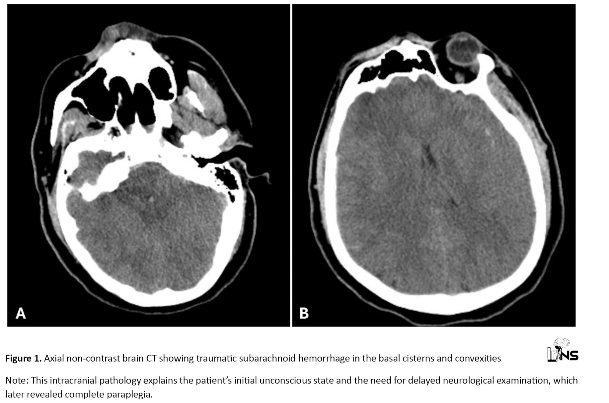
A 25-year-old male was admitted to the emergency department after being involved in a high-energy motor vehicle accident. On arrival, he was unconscious and exhibited signs of polytrauma, including multiple contusions and suspected head injury (Figure 1).
 Endotracheal intubation was performed under sedation due to agitation and compromised airway protection.
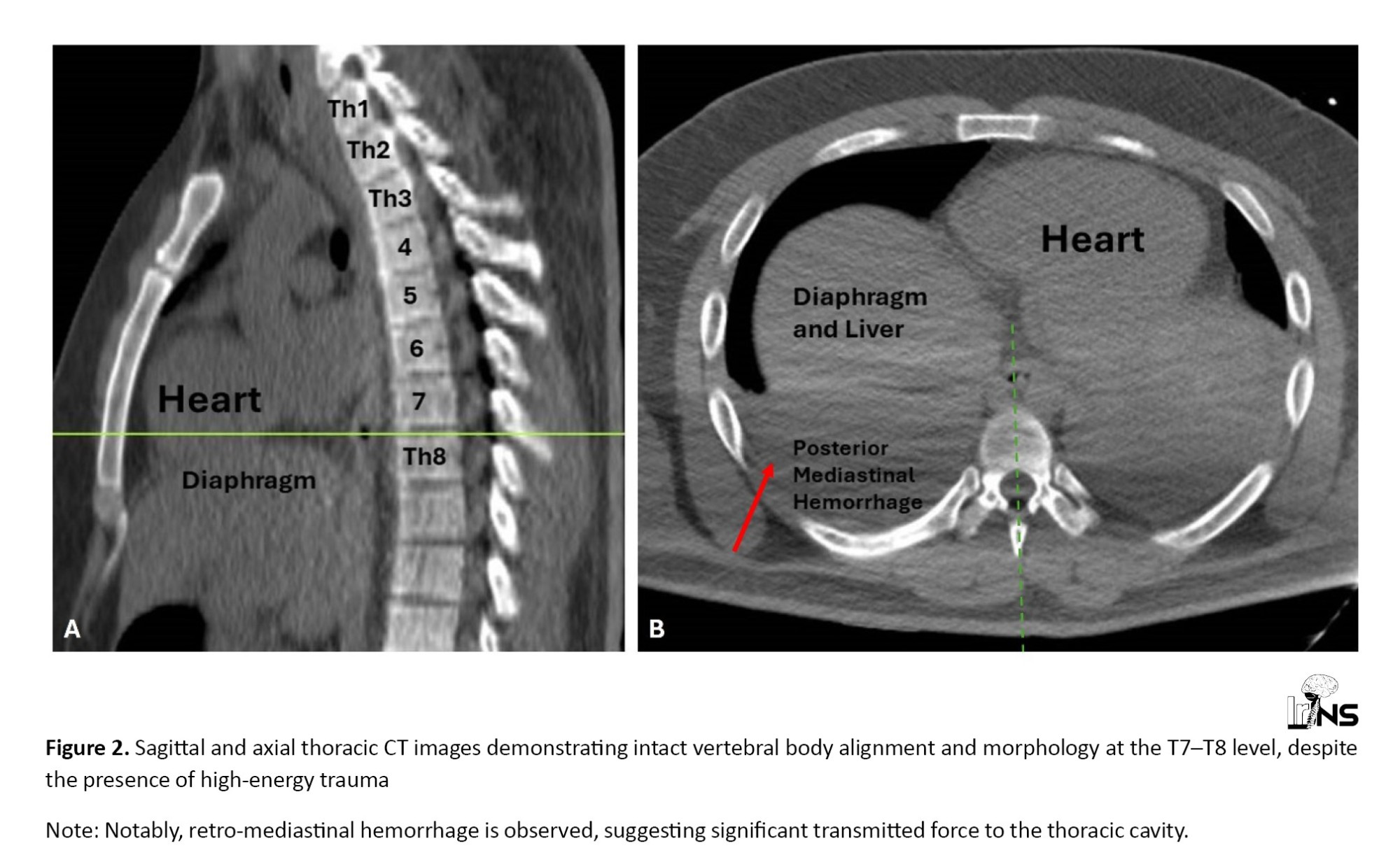
Initial brain computerized tomography CT revealed a traumatic subarachnoid hemorrhage. Thoracic CT showed right-sided rib fractures at the 7th and 8th levels, left-sided rib fractures at the second, fourth, and ninth levels, bilateral pleural effusion, and pulmonary contusions. However, no evidence of thoracic vertebral fracture, subluxation, or dislocation was observed. The patient was admitted to the intensive care unit and kept under sedation for 48 hours due to the severity of his cranial trauma.
Upon regaining consciousness one week later, neurological examination revealed a Glasgow Coma scale (GCS) score of 15, but complete motor paralysis of both lower extremities was noted. Deep tendon reflexes were absent in the legs, and anal sphincter tone was diminished. Sensory examination demonstrated a sharp sensory level at T7. There was no voluntary movement or proprioception below this level, consistent with complete paraplegia. The thoracic CT images were re-evaluated and again confirmed the absence of bony injury (Figure 2).
Radiological findings
A thoracic magnetic resonance imaging (MRI) scan was obtained to investigate the cause of the neurologic deficit. T2-weighted sagittal and axial images revealed a complete transection of the spinal cord at the T7–T8 level (Figure 3).
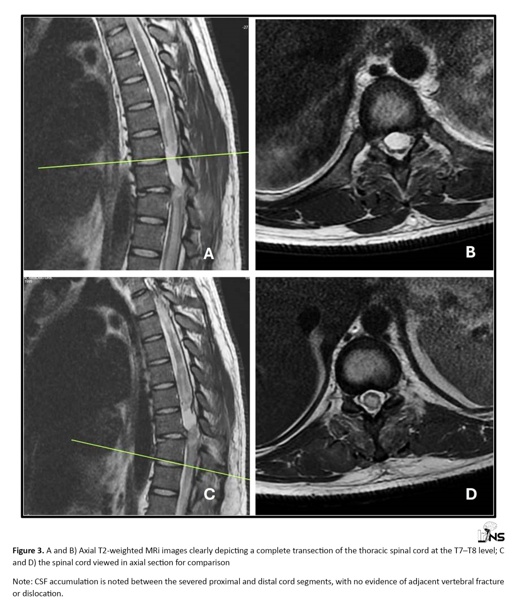 The cord was visibly disrupted, with cerebrospinal fluid (CSF) clearly delineating the gap between the proximal and distal stumps. No signs of vertebral body fracture, facet joint dislocation, or posterior element disruption were seen.
There were also no signs of epidural hematoma, ligamentous injury, or spinal instability. Importantly, the alignment of the vertebral column was preserved, and the intervertebral disc spaces were intact. These findings supported the diagnosis of a complete SCIWORA, in its rare thoracic adult form. Given the biomechanical stability of the spinal column and absence of compressive pathology, surgical intervention was not pursued, and the patient was managed conservatively.
3. Discussion
SCIWORA was originally conceptualized to describe spinal cord injury in pediatric patients who presented with neurologic deficits following trauma but lacked evidence of osseous injury on radiographs or CT scans [1]. This phenomenon is most frequently observed in children due to their flexible ligamentous structures and immature musculoskeletal system. In contrast, adult SCIWORA is rare, particularly in the thoracic region, where the spine is anatomically more rigid and stabilized by the rib cage [4, 5].
The presented case illustrates an unusual form of adult thoracic SCIWORA, in which a complete transection of the spinal cord occurred without any radiological evidence of fracture, dislocation, or biomechanical instability. The rarity of such presentations is supported by existing literature: A review by Khatri et al. identified only a handful of thoracic SCIWORA cases in adults, most of which involved edema or signal change, rather than frank cord transection [6]. Cases of MRI-confirmed complete transection in the absence of vertebral injury are exceptionally uncommon [7–9].
Several injury mechanisms have been proposed for such presentations, including hyperflexion or hyperextension forces that cause distraction injuries to the cord without bone injury, vascular compromise leading to ischemia and secondary cord necrosis, and shearing forces transmitted through intact vertebral columns in high-energy trauma [10–13].
These rare cases highlight a potential gap in the current classification of spinal cord injuries. The term SCIWORA, though useful, fails to adequately capture the full range of presentations, especially in adults, where high-resolution MRI has demonstrated significant parenchymal injury despite normal radiographs and CT imaging. Some authors have proposed alternate terminology, such as “SCIWOCTET” or “SCIWOBA,” to reflect this broader spectrum [14]. However, these terms remain inconsistently used and lack formal validation.
A review of previously published adult thoracic SCIWORA cases is summarized in Table 1.
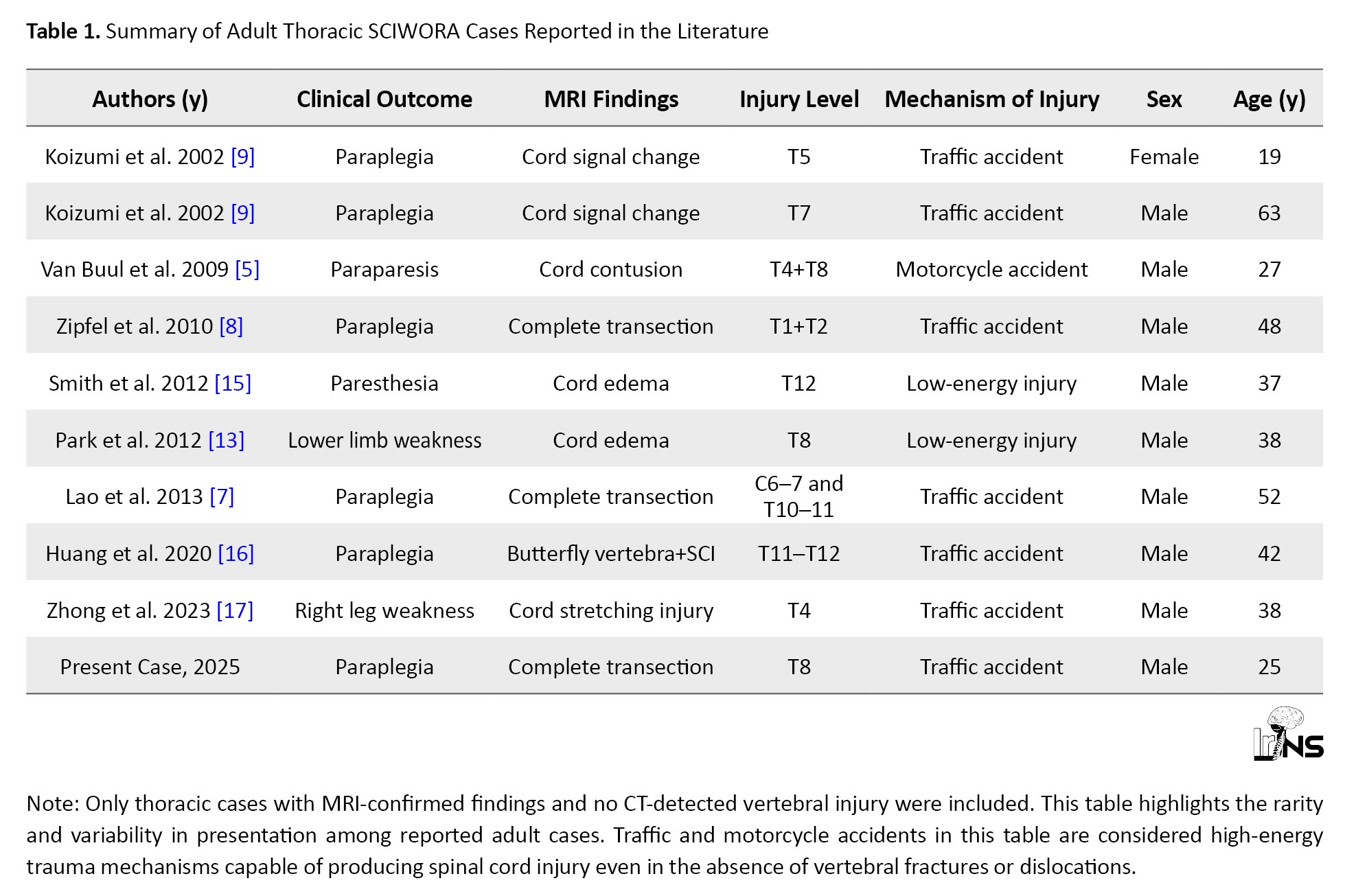
As demonstrated, complete spinal cord transection in this context is extremely rare, with only a few reported cases in the literature, underscoring the exceptional nature of our case.
Our case supports the need for a refined classification framework that recognizes the radiological-histopathological disconnect in adult thoracic injuries, accounts for MRI-only detectable injuries, differentiates between minor cord edema and complete transection, and includes a biomechanical assessment component.
Such a system may aid in prognostication, guide therapeutic decision-making, and standardize reporting in clinical and academic settings.
Delayed diagnosis and preventive strategies
In retrospect, this case raises the critical issue of diagnostic delay in spinal cord injuries without radiographic abnormalities. Although the patient was sedated due to severe cranial trauma, a comprehensive spinal examination might have prompted earlier suspicion of spinal cord injury, especially in the presence of high-energy trauma and associated thoracic injuries such as bilateral rib fractures and pulmonary contusions. Early neurological assessment, including passive examination of deep tendon reflexes or sensory evaluation where feasible, could have triggered earlier imaging.
Similar diagnostic delays have been reported in the literature. In a case reported by Dubey et al., an adult patient developed paraplegia several hours after trauma, with the initial CT being normal and an MRI performed only after neurological deterioration [12]. Likewise, Hana et al. described a case of delayed-onset paraplegia in which MRI findings consistent with SCIWORA were only detected after a 24-hour lag, contributing to poorer clinical outcomes [18]. These reports, along with our case, underscore the need for high suspicion in trauma patients with altered consciousness or distracting injuries.
To minimize diagnostic delay, current recommendations suggest that MRI should be performed in any trauma patient with unexplained neurological findings, particularly when CT and X-rays are unremarkable. This approach is supported by recent guidelines in spinal trauma care, which advocate for early MRI in cases of neurologic deficit despite normal radiographs [4, 13].
4. Conclusion
Thoracic SCIWORA in adults remains an exceptionally rare entity, particularly when involving complete cord transection without vertebral fracture or dislocation. This case underscores the critical importance of early MRI in patients presenting with severe neurological deficits and unremarkable initial radiographs or CT scans. The findings support the existence of a distinct subgroup of adult thoracic SCIWORA that current classification systems may not fully capture. There is a need to revisit and potentially refine the terminology and diagnostic criteria to incorporate MRI-based findings and biomechanical assessments, thereby improving recognition, communication, and management strategies in such complex cases.
Ethical Considerations
Compliance with ethical guidelines
This case report was conducted in accordance with institutional and international ethical standards. Informed consent was obtained from the patient for publication of all clinical information and accompanying images. Ethical approval was not required for a single case report under local regulations.
Funding
This research did not receive any grant from funding agencies in the public, commercial, or non-profit sectors.
Authors' contributions
Conceptualization and study design: Vaner Köksal and Cem Demirel; Data collection: Melih Van, and Fatih Tomakin; Data analysis and interpretation: Melih Van; Final approval: Vaner Köksal; Review & editing: Vaner Köksal and Recai Engin.
Conflict of interest
The authors declared no conflict of interest.
References
Spinal cord injury without radiographic abnormality (SCIWORA) was first introduced by Pang and Wilberger in 1982 to describe spinal cord injuries in pediatric patients whose radiographs and computed tomography (CT) scans showed no signs of vertebral fracture or dislocation [1]. The term has historically been associated with the pediatric population, particularly in relation to the cervical spine, due to the greater elasticity and flexibility of the immature spinal column [2].
With the advent of magnetic resonance imaging (MRI), our ability to detect intramedullary spinal cord damage has significantly improved, leading to the identification of SCIWORA-like presentations in adult patients as well [3]. However, in contrast to pediatric cases, adult cases of SCIWORA are rare and often debated due to the relative rigidity of the mature spinal column and reduced ligamentous laxity [4, 5].
The thoracic spine, being the most rigid and biomechanically stable segment of the vertebral column, is especially resistant to injury without associated osseous or ligamentous disruption. Consequently, thoracic SCIWORA in adults is exceedingly uncommon, with only a limited number of reported cases in the literature [6–9].
Herein, we present a rare case of an adult patient who developed complete paraplegia following a high-energy motor vehicle accident, in the absence of any thoracic vertebral fracture or dislocation. MRI subsequently revealed a full-thickness transection of the spinal cord at the T7–T8 level. This case underscores the importance of maintaining a high index of suspicion for spinal cord injury in trauma patients with neurological deficits but unremarkable initial radiographs or CT scans. It also contributes to the limited body of literature documenting thoracic SCIWORA in skeletally mature individuals.
2. Case Presentation
A 25-year-old male was admitted to the emergency department after being involved in a high-energy motor vehicle accident. On arrival, he was unconscious and exhibited signs of polytrauma, including multiple contusions and suspected head injury (Figure 1).
Initial brain computerized tomography CT revealed a traumatic subarachnoid hemorrhage. Thoracic CT showed right-sided rib fractures at the 7th and 8th levels, left-sided rib fractures at the second, fourth, and ninth levels, bilateral pleural effusion, and pulmonary contusions. However, no evidence of thoracic vertebral fracture, subluxation, or dislocation was observed. The patient was admitted to the intensive care unit and kept under sedation for 48 hours due to the severity of his cranial trauma.
Upon regaining consciousness one week later, neurological examination revealed a Glasgow Coma scale (GCS) score of 15, but complete motor paralysis of both lower extremities was noted. Deep tendon reflexes were absent in the legs, and anal sphincter tone was diminished. Sensory examination demonstrated a sharp sensory level at T7. There was no voluntary movement or proprioception below this level, consistent with complete paraplegia. The thoracic CT images were re-evaluated and again confirmed the absence of bony injury (Figure 2).
Radiological findings
A thoracic magnetic resonance imaging (MRI) scan was obtained to investigate the cause of the neurologic deficit. T2-weighted sagittal and axial images revealed a complete transection of the spinal cord at the T7–T8 level (Figure 3).
There were also no signs of epidural hematoma, ligamentous injury, or spinal instability. Importantly, the alignment of the vertebral column was preserved, and the intervertebral disc spaces were intact. These findings supported the diagnosis of a complete SCIWORA, in its rare thoracic adult form. Given the biomechanical stability of the spinal column and absence of compressive pathology, surgical intervention was not pursued, and the patient was managed conservatively.
3. Discussion
SCIWORA was originally conceptualized to describe spinal cord injury in pediatric patients who presented with neurologic deficits following trauma but lacked evidence of osseous injury on radiographs or CT scans [1]. This phenomenon is most frequently observed in children due to their flexible ligamentous structures and immature musculoskeletal system. In contrast, adult SCIWORA is rare, particularly in the thoracic region, where the spine is anatomically more rigid and stabilized by the rib cage [4, 5].
The presented case illustrates an unusual form of adult thoracic SCIWORA, in which a complete transection of the spinal cord occurred without any radiological evidence of fracture, dislocation, or biomechanical instability. The rarity of such presentations is supported by existing literature: A review by Khatri et al. identified only a handful of thoracic SCIWORA cases in adults, most of which involved edema or signal change, rather than frank cord transection [6]. Cases of MRI-confirmed complete transection in the absence of vertebral injury are exceptionally uncommon [7–9].
Several injury mechanisms have been proposed for such presentations, including hyperflexion or hyperextension forces that cause distraction injuries to the cord without bone injury, vascular compromise leading to ischemia and secondary cord necrosis, and shearing forces transmitted through intact vertebral columns in high-energy trauma [10–13].
These rare cases highlight a potential gap in the current classification of spinal cord injuries. The term SCIWORA, though useful, fails to adequately capture the full range of presentations, especially in adults, where high-resolution MRI has demonstrated significant parenchymal injury despite normal radiographs and CT imaging. Some authors have proposed alternate terminology, such as “SCIWOCTET” or “SCIWOBA,” to reflect this broader spectrum [14]. However, these terms remain inconsistently used and lack formal validation.
A review of previously published adult thoracic SCIWORA cases is summarized in Table 1.
As demonstrated, complete spinal cord transection in this context is extremely rare, with only a few reported cases in the literature, underscoring the exceptional nature of our case.
Our case supports the need for a refined classification framework that recognizes the radiological-histopathological disconnect in adult thoracic injuries, accounts for MRI-only detectable injuries, differentiates between minor cord edema and complete transection, and includes a biomechanical assessment component.
Such a system may aid in prognostication, guide therapeutic decision-making, and standardize reporting in clinical and academic settings.
Delayed diagnosis and preventive strategies
In retrospect, this case raises the critical issue of diagnostic delay in spinal cord injuries without radiographic abnormalities. Although the patient was sedated due to severe cranial trauma, a comprehensive spinal examination might have prompted earlier suspicion of spinal cord injury, especially in the presence of high-energy trauma and associated thoracic injuries such as bilateral rib fractures and pulmonary contusions. Early neurological assessment, including passive examination of deep tendon reflexes or sensory evaluation where feasible, could have triggered earlier imaging.
Similar diagnostic delays have been reported in the literature. In a case reported by Dubey et al., an adult patient developed paraplegia several hours after trauma, with the initial CT being normal and an MRI performed only after neurological deterioration [12]. Likewise, Hana et al. described a case of delayed-onset paraplegia in which MRI findings consistent with SCIWORA were only detected after a 24-hour lag, contributing to poorer clinical outcomes [18]. These reports, along with our case, underscore the need for high suspicion in trauma patients with altered consciousness or distracting injuries.
To minimize diagnostic delay, current recommendations suggest that MRI should be performed in any trauma patient with unexplained neurological findings, particularly when CT and X-rays are unremarkable. This approach is supported by recent guidelines in spinal trauma care, which advocate for early MRI in cases of neurologic deficit despite normal radiographs [4, 13].
4. Conclusion
Thoracic SCIWORA in adults remains an exceptionally rare entity, particularly when involving complete cord transection without vertebral fracture or dislocation. This case underscores the critical importance of early MRI in patients presenting with severe neurological deficits and unremarkable initial radiographs or CT scans. The findings support the existence of a distinct subgroup of adult thoracic SCIWORA that current classification systems may not fully capture. There is a need to revisit and potentially refine the terminology and diagnostic criteria to incorporate MRI-based findings and biomechanical assessments, thereby improving recognition, communication, and management strategies in such complex cases.
Ethical Considerations
Compliance with ethical guidelines
This case report was conducted in accordance with institutional and international ethical standards. Informed consent was obtained from the patient for publication of all clinical information and accompanying images. Ethical approval was not required for a single case report under local regulations.
Funding
This research did not receive any grant from funding agencies in the public, commercial, or non-profit sectors.
Authors' contributions
Conceptualization and study design: Vaner Köksal and Cem Demirel; Data collection: Melih Van, and Fatih Tomakin; Data analysis and interpretation: Melih Van; Final approval: Vaner Köksal; Review & editing: Vaner Köksal and Recai Engin.
Conflict of interest
The authors declared no conflict of interest.
References
- Pang D, Wilberger JE Jr. Spinal cord injury without radiographic abnormalities in children. Journal of Neurosurgery. 1982; 57(1):114-29. [DOI:10.3171/jns.1982.57.1.0114] [PMID]
- Rosche C, Carty H. Spinal trauma in children. Pediatric Radiology. 2001; 31(9):677-700. [DOI:10.1007/s002470100532] [PMID]
- Martinez-Perez R, Joswig H, Rayo N, Bertazzo-Silveira G. Spinal cord injury without radiological abnormalities and the importance of magnetic resonance. Acta Bio Medica. 2020; 91(3):e2020084. [DOI:10.23750/abm.v91i3.8518] [PMID]
- Hirsh LF, Duarte L, Wolfson EH. Thoracic spinal cord injury without spine fracture in an adult: Case report and literature review. Surgical Neurology. 1993; 40(1):35-8. [DOI:10.1016/0090-3019(93)90167-Y] [PMID]
- Van Buul G, Oner FC. Thoracic spinal cord injury without radiographic abnormality in an adult patient. The Spine Journal. 2009; 9(3):e5-8. [DOI:10.1016/j.spinee.2008.03.003] [PMID]
- Khatri K, Farooque K, Gupta A, Sharma V. Spinal cord injury without radiological abnormality in adult thoracic spinal trauma. Archives of Trauma Research. 2014; 3(3):e19036. [DOI:10.5812/atr.19036] [PMID]
- Lao LF, Zhong GB, Liu ZD. Transection of double-level spinal cord without radiographic abnormalities in an adult: A case report. Orthopaedic Surgery. 2013; 5(4):302-4. [DOI:10.1111/os.12065] [PMID]
- Zipfel B, Buz S, Hullmeine D, Röttgen R, Hetzer R. Traumatic transection of the aorta and thoracic spinal cord injury without radiographic abnormality in an adult patient. Journal of Endovascular Therapy. 2010; 17(1):131-6. [DOI:10.1583/09-2837MR-R.1] [PMID]
- Koizumi M, Ueda Y, Iida J, Matsuyama E, Yoshikawa T, Takakura Y, et al. Upper thoracic spinal cord injury without vertebral bony lesion: A report of two cases. Spine. 2002; 27(21):E467-70. [DOI:10.1097/00007632-200211010-00020] [PMID]
- Schmidt P, Mayer TE, Drescher R. Delineation of alar ligament morphology: Comparison of magnetic resonance imaging at 1.5 and 3 Tesla. Orthopedics. 2012; 35(11):e1635-9. [DOI:10.3928/01477447-20121023-22] [PMID]
- Gupta SK, Rajeev K, Khosla VK, Sharma BS, Paramjit, Mathuriya SN, et al. Spinal cord injury without radiographic abnormality in adults. Spinal Cord. 1999; 37(10):726-9. [DOI:10.1038/sj.sc.3100900] [PMID]
- Dubey A, Tomar S, Gupta A, Khandelwal D. Delayed paraplegia in an adult patient with spinal cord injury without radiographic abnormality of dorsal spine: A lesson learned. Asian Journal of Neurosurgery. 2018; 13(3):867-9. [DOI:10.4103/1793-5482.238013] [PMID]
- Park MC, Bok SK, Lee SJ, Ahn DH, Lee YJ. Delayed onset of thoracic SCIWORA in adults. Annals of Rehabilitation Medicine. 2012; 36(6):871-5. [DOI:10.5535/arm.2012.36.6.871] [PMID]
- Schlenk RP, Kowalski RJ, Benzel EC. Biomechanics of spinal deformity. Neurosurgical Focus. 2003; 14(1):e2. [DOI:10.3171/foc.2003.14.1.3] [PMID]
- Smith HK, Durnford AJ, Sherlala K, Merriam WF. Lower thoracic spinal cord injury without radiographic abnormality in an amateur rugby player. BMJ Case Reports. 2012; 2012:bcr0120125716. [DOI:10.1136/bcr.01.2012.5716] [PMID]
- Huang AB, Bai M, Liu H, Zhou ZY, Wei JX. Spinal cord injury in an adult patient with thoracic butterfly vertebra: A case report and review of the literature. BMC Musculoskeletal Disorders. 2020; 21(1):392. [DOI:10.1186/s12891-020-03433-9] [PMID]
- Zhong W, Pu M, Zhong Y, Zhong P, Shen Y. Spinal cord injury without radiographic abnormalities caused by rotation stretching injury manifesting as brown- sequard syndrome: A case report. The Journal of the Pakistan Medical Association. 2023; 73(8):1732-4. [DOI:10.47391/JPMA.7779] [PMID]
- Hana H, Gnena H, Safia O, Rym BK, Jendoubi A, Aymen Z, et al. Delayed presentation of spinal cord injury without radiographic abnormality (SCIWORA): A case report. The Pan African Medical Journal. 2023; 45:160. [DOI:10.11604/pamj.2023.45.160.23467] [PMID]
Type of Study: Case report |
Subject:
Spine
| Rights and Permissions | |
 |
This work is licensed under a Creative Commons Attribution-NonCommercial 4.0 International License. |

Copyright © The Author(s);
This is an open access article distributed under the terms of the Creative Commons Attribution License (CC-By-NC), which permits use, distribution, and reproduction in any medium, provided the original work is properly cited and is not used for commercial purposes.
Contact Information
This is an open access article distributed under the terms of the Creative Commons Attribution License (CC-By-NC), which permits use, distribution, and reproduction in any medium, provided the original work is properly cited and is not used for commercial purposes.
Contact Information
