Sun, Jul 26, 2026
Volume 11 - Continuous Publishing
Iran J Neurosurg 2025, 11 - Continuous Publishing: 0-0 |
Back to browse issues page


Download citation:
BibTeX | RIS | EndNote | Medlars | ProCite | Reference Manager | RefWorks
Send citation to:


BibTeX | RIS | EndNote | Medlars | ProCite | Reference Manager | RefWorks
Send citation to:
Doostabadi H, Esmaeili F. Comparison of neuro-endoscopic and non-endoscopic surgical techniques in the treatment of septated chronic subdural hematoma.. Iran J Neurosurg 2025; 11 : 27
URL: http://irjns.org/article-1-483-en.html
URL: http://irjns.org/article-1-483-en.html


1- Departments of Neurosurgery, School of Medicine, Hamadan University of Medical Sciences, Hamadan, Iran.
Full Text [PDF 840 kb]
(128 Downloads)
| Abstract (HTML) (458 Views)
Full Text: (63 Views)
1. Introduction
Chronic subdural hematoma (CSDH) usually occurs in the sixth and seventh decades of life. It refers to the presence of a dural tear (for at least 20 days) between the dura mater and the arachnoid in the brain. CSDH is clinically important because it can lead to severe permanent complications and even death [1]. A prevalent condition, CSDH occur 39 times per 100000 persons annually, with 45 incidences per 100000 people between the ages of 70 and 79 [2, 3]. Symptoms of the disease include headache, memory impairment, confusion, etc [4]. CSDH is usually attributed to a history of head trauma (even minor and low-power trauma that the patient himself does not remember) [5]. Septated CSDH (sCSDH) is a special type of CSDH characterized by a hematoma cavity, divided into several smaller cavities by multiple fibrous septa. CSDH formation may result from damage to the septa in the bridging vessels (which run from the brain to the draining veins), which usually becomes symptomatic 4 to 7 weeks after the trauma [6-8].
Today, the most suitable treatment for this complication has not been established, and although cases of spontaneous hematoma absorption have been reported, the best treatment remains surgery [9, 10].
Several surgical approaches have been employed to manage CSDHs. One of the oldest methods is craniotomy, which is used when a solid component is present in the hematoma, the brain is not expanded after the hematoma is drained, or when there is relapse [11]. The twist-drill trephination method can also be performed on the patient’s bed, especially for sick patients under local anesthesia. The highest risk of recurrence is related to this method [12]. Today, the most widely utilized procedure is burr hole craniostomy with or without drainage. The recurrence rate is reported to be 3-35%, is associated with more complications, and yields similar results [13].
Chronic subdural septate hematoma is a significant issue in modern neurosurgery because a simple open hole is not usually enough to drain the hematoma. Actually, a larger craniotomy and membrane resection are required to drain the hematoma, which is associated with an elevated degree of mortality and morbidity [14]. The endoscopic method provides the surgeon with direct vision during the operation. It allows the surgeon to visualize the septa in the bridging veins and remove them safely if needed, thereby greatly reducing the risk of recurrence. However, there is insufficient visibility during nonendoscopic drainage of CSDH, especially in multi-cavity hematomas. So, surgeons have not yet reached a settled, reliable standard of care for the treatment of CSDH. The purpose of this study is to compare two neuroendoscopic and nonendoscopic surgical approaches used in the therapeutic process of CSDH with septa.
2. Methods and Materials
Trial design
Our study population was the patients referred to the Hamadan Research and Educational Hospital. This study was submitted to the Ethics Committee of Hamadan University of Medical Sciences, and the ethics code number was taken. It was registered on the Iranian clinical trial site and confirmed. All stages of this research were done with permission from the relevant authorities. All checklists used were anonymous and based on numbers, and participants received assurances that their information would remain confidential within the research group.
Study participants
All participants were fully informed, and their written informed consent was obtained. In this clinical trial, 32 patients were randomly assigned to two groups using the simple random block method: One group received endoscopic and the other, open techniques. This randomized controlled trial compared neuroendoscopic surgery (n=16) with conventional burr-hole craniostomy (n=16) in patients with septated CSDH. The patients were informed that participation in this research is voluntary and that they may not receive services if they choose not to participate. The treatment at that center will not be available for them. The inclusion criteria for the study were as follows: patients with CSDH, patients with mixed-density computed tomography (CT), and patients or their companions who consented to participate in the study. The exclusion criteria were as follows: patients with a low level of consciousness (Glasgow coma scale [GCS] less than 9), acute subdural hematoma, patients with involvement of several organs, patients with a history of liver cirrhosis and coagulation disorders, and patients with a history of shunt.
Interventions (surgical techniques)
CSDH shows a wide range of symptoms. All emergency patients or patients diagnosed with CT in the clinic were involved in the research. The presence of a septum was determined using a CT scan. Septation in CT was determined by the presence of layers separating the hematoma. To control for bias, all surgical procedures were performed by a single surgeon to minimize technical variability after a complete examination and detailed history (including changes in age, gender, blood pressure, coagulant intake, and level of consciousness, as assessed by GCS). They were separated into endoscopic and nonendoscopic groups. In the control group, the barhol method (two barhols) was performed in the supine position after the patient was anesthetized in the operating room. In this method, after a 5-cm skin incision (according to the location of the hematoma), the skull was drilled with two drills (piercing the skull to the dura mater surface). Given the hematoma’s size and location, a 3-cm hole was created. The dura was burned with bipolar, and careful blood sampling was done to prevent blood from entering the brain. The edges of the dura mater were attached to the bone wall with bipolar or cautery, and at this stage, the brain cortex was visible. At the end, we washed with plenty of warm serum, achieved sufficient hemostasis, and, after clearing, placed a drain and sutured the incision site. In the endoscopy method, after preparing the endoscope, a skin incision was made similar to the previous method; however, an S-shaped incision was used to facilitate endoscope use. The borehole was placed in the right place, and after creating a hole in the skull, we used a cresson punch to enlarge it (about 3-5 cm). After draining the hematoma and washing, we entered the subdural space with an endoscope (zero-degree rigid) and examined the vascular trabeculae or septa under direct vision. If a vascular trabecula was seen, it was burned in the same place with a bipolar endoscope. Also, if there were septa, the septa were opened one by one and washed to prevent recurrence.
At the same time, the control group was drained and sutured after hemostasis was ensured. After the operation, the procedure duration was compared between the two groups. Also, they were followed up for 6 months, and mortality, morbidity, and other complications were carefully examined and compared according to Markwalder criteria (Table 1).
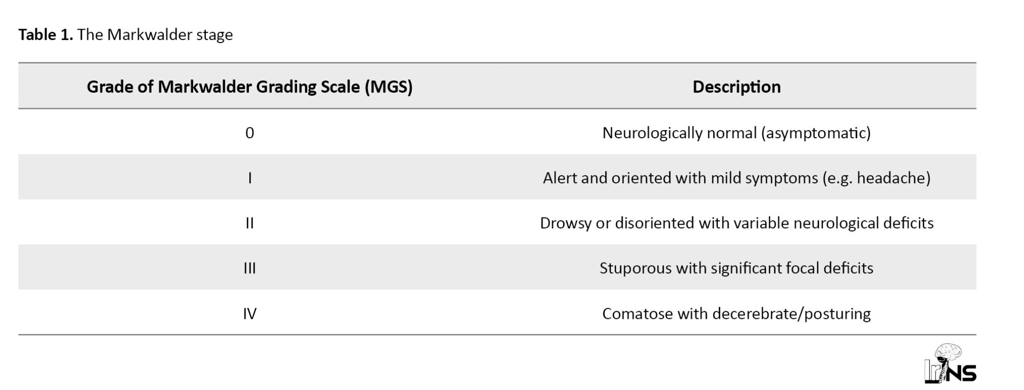
This criterion was divided into 0-IV based on the level of consciousness and the presence of neurological deficits. In addition, to avoid bias, the same surgeon conducted both operations.
Statistical analysis
Data analysis was performed using SPSS software, version 22. Frequency and percentage were used to characterize qualitative variables. The Mean±SD were used to characterize quantitative variables. The independent t-test (age parameter) or the Mann-Whitney test (bleeding rate parameter, duration of hospitalization, and duration of surgery) was used to compare the averages between the two groups. To compare qualitative data between groups, the chi-square test was used. The Kolmogorov-Smirnov test was used to determine whether the data followed a normal distribution. The significance level was considered 0.05 in all cases.
3. Results
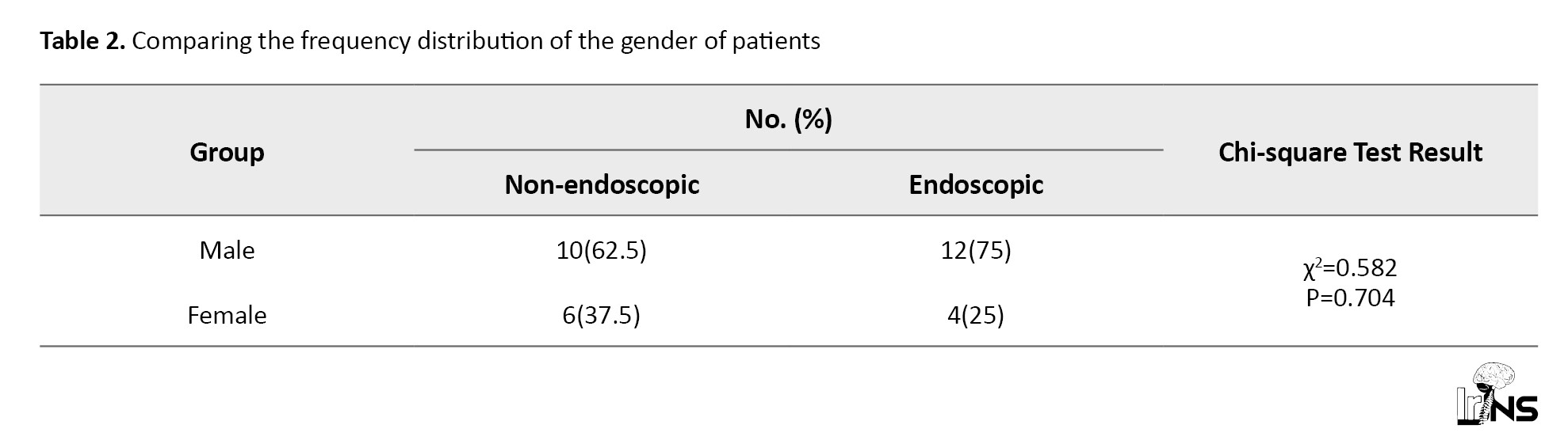
In the present study, 32 patients referred to the emergency room of Besat Hospital in Hamadan were examined—16 in the nonendoscopic group and 16 in the endoscopic surgery group. The findings of the current research indicate no significant difference in average age or gender between the non-endoscopy and endoscopic groups (P>0.05).
Based on the findings of the present study in Tables 2, and 3, the frequency distribution of clinical symptoms, GCS level, and history of trauma in the two groups of nonendoscopic and endoscopic patients was not significantly different from each other (P>0.05).
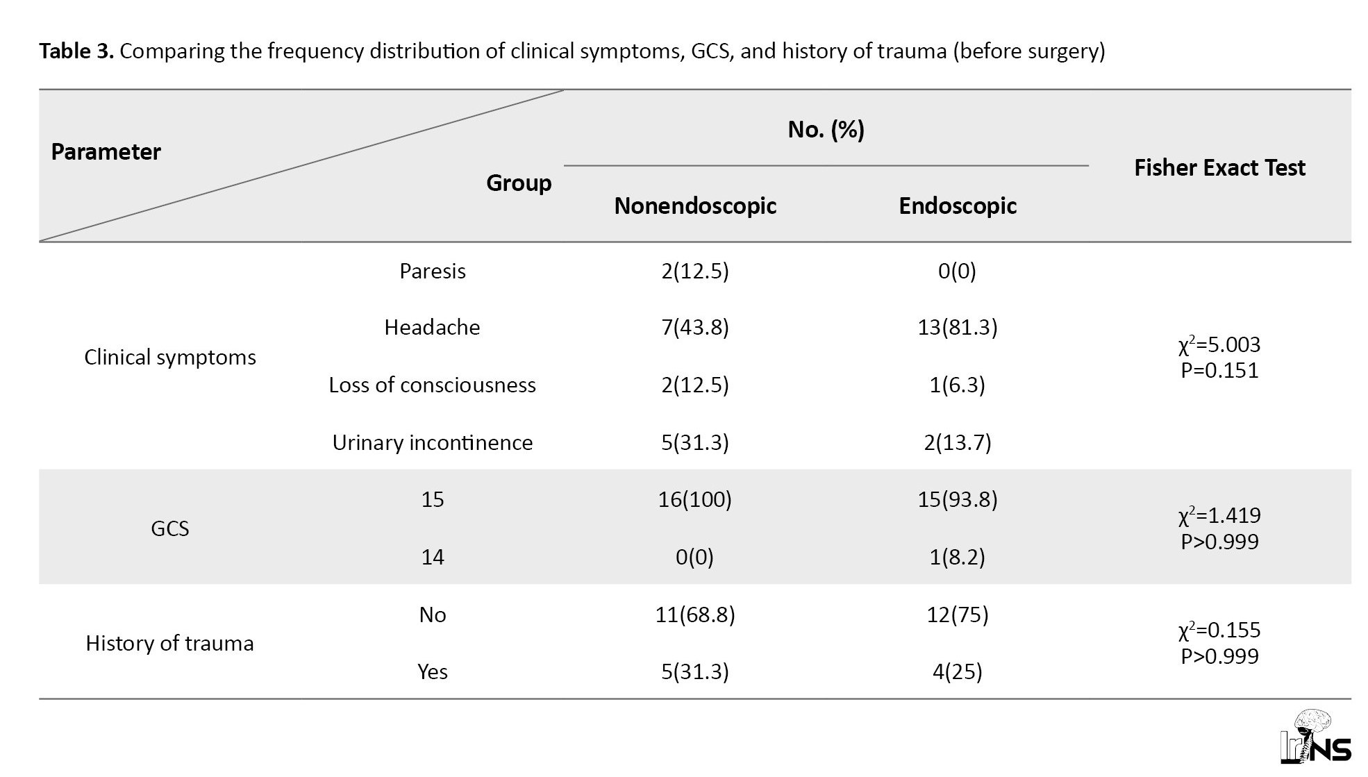
According to the results of the present study (Table 3), the mean hospitalization duration and bleeding in the endoscopic group were significantly reduced compared to the nonendoscopic group (P<0.05). Also, despite the longer surgical duration, there was no significant difference in the endoscopic group compared with the nonendoscopic group (P=0.086).
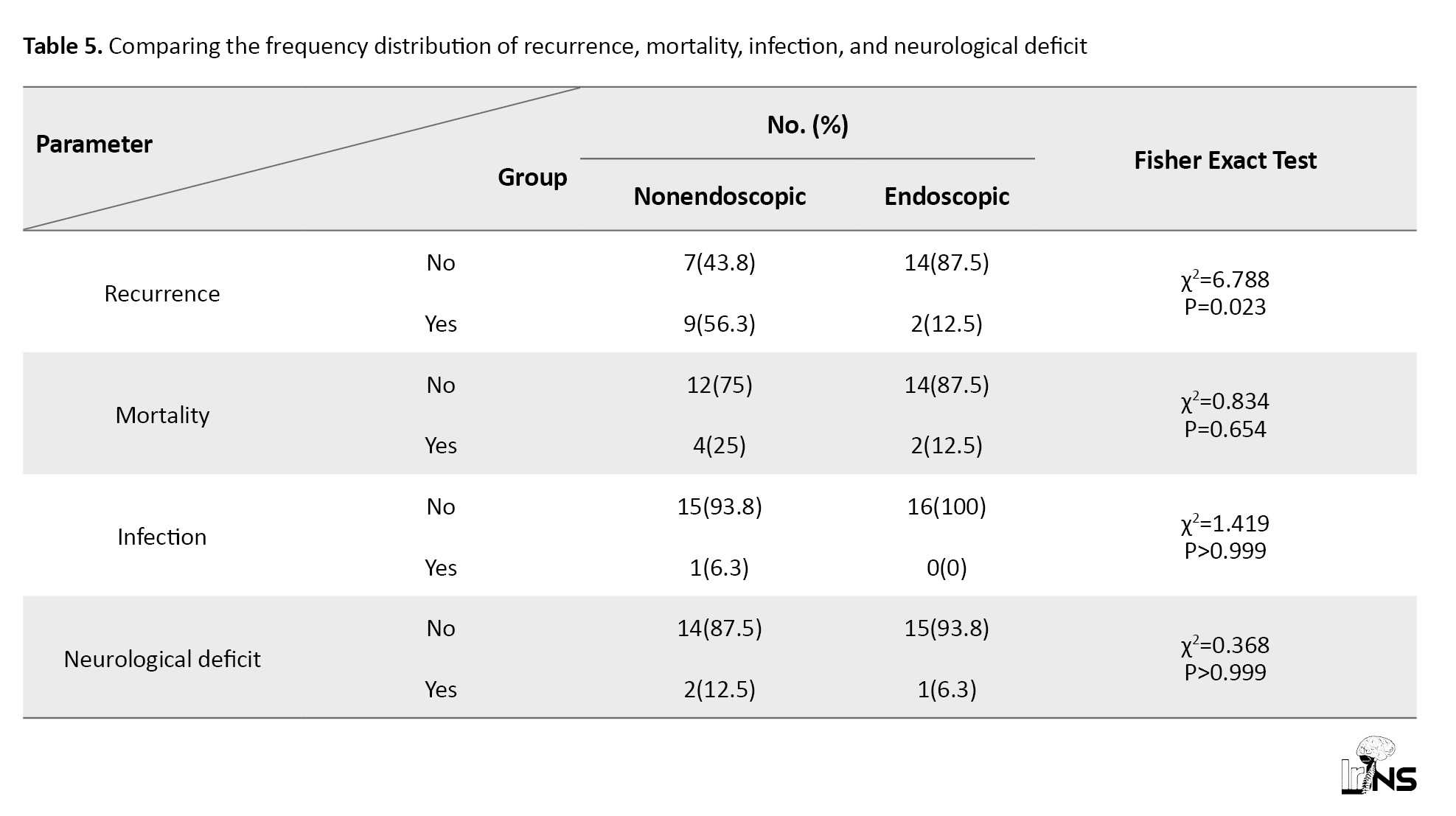
Based on the findings of the present study (Table 4), the frequency of death, neurological complications, and infection (within 6 months) was lower in the endoscopic group than in the other group.
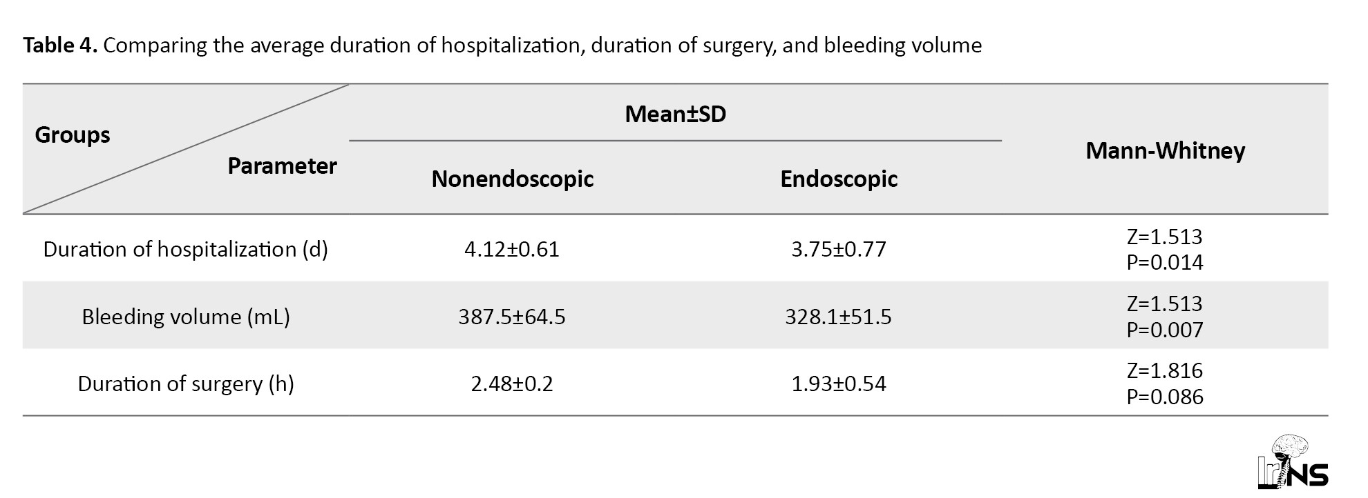
However, this difference was not significant (P>0.05). Also, no hematoma was observed in any patient in either group. Also, the recurrence frequency distribution in the endoscopic group was significantly lower than in the nonendoscopic group (P=0.023).
4. Discussion
According to the results of the present study, all patients were hospitalized in the intensive care unit (ICU) for 1 day after admission and then transferred to the ward. In the study by Abrishamkar et al., the average ICU hospitalization duration after surgery showed no significant difference between the two groups, consistent with our study [15]. According to the current study, patients in the endoscopy group experienced significantly less bleeding and a shorter average length of hospitalization than those in the non-endoscopy group (P<0.05). The duration of surgery did not differ significantly between the two groups (P=0.086). In a study by Abrishamkar et al., the average hospitalization and bleeding durations of patients undergoing neuroendoscopic surgery were significantly shorter than those of the nonendoscopic group, consistent with our study [15]. Also, in the study by Du et al., the average surgical duration in the endoscopic group was significantly longer than in the nonendoscopic group, which was inconsistent with our study [16]. Among the reasons for this controversy, we can mention differences in the timing of the study, the availability of facilities and equipment, and the surgeons’ experience. According to the Guan et al. study, there was no significant difference in the average surgical duration between the endoscopic and nonendoscopic groups, consistent with our study [17].
Considering the findings of the present study, there was no significant difference in the frequency distribution of trauma, mortality, neurological complications, and infection in the nonendoscopic and endoscopic groups of patients (P>0.05). Also, compared to the nonendoscopic group, the endoscopic group’s recurrence frequency distribution was significantly lower (P=0.023). According to the investigation by Allahdini et al., the recurrence rate in the endoscopic and nonendoscopic surgery groups did not differ significantly, which was inconsistent with the present study [18]. Taussky et al. reported that the recurrence rate with the nonendoscopic method was significantly lower than with the endoscopic method, which was inconsistent with our study [19]. Among the reasons for this inconsistency, we can mention differences in the study population and the study period.
Du et al. testified that the recurrence rate in endoscopic surgery patients was lower than in the nonendoscopic group [16]. Guan et al. reported that the recurrence rate after endoscopic surgery was much lower than in the nonendoscopic group, consistent with our study [17].
Ishikawa et al. reported that during follow-up after endoscopic surgery, all patients’ neurological function improved, and no neurological defects were observed [20]. In the study by Ichimura et al., no hematoma recurrence was observed 6 months after surgery in any patient, which was inconsistent with our study [21].
Abrishamkar et al. reported that the distribution of death frequencies between the two investigated groups was not significantly different, consistent with our study [15].
It seems that the follow-up of subdural hematoma patients for 6 months after surgery is not necessary unless there are risk factors such as high or mixed density in the CT scan, taking antiplatelet drugs, or other concomitant diseases in the patient. In accordance with a different study by Javadi et al., patients with CSDHs do not require follow-up beyond one month [22].
5. Conclusion
In this study, we examined endoscopic and nonendoscopic methods in the treatment of CSDH. Except for the operation time, which was slightly higher in the endoscopic group (not significant) than in the non-endoscope group, in other cases, including infection, neurological deficit, bleeding rate, and mortality, it was lower in the endoscope group. Also, the recurrence rate with the endoscopic method is lower than with the nonendoscopic method (significant). Therefore, the endoscopic method can be considered the preferred approach for the surgical treatment of CSDH with septa.
Ethical Considerations
Compliance with ethical guidelines
This study was registered by the Iranian Registry of Clinical Trials (IRCT), Tehran, Iran (CODE: IRCT20190618043934N14). This study was approved by the Research Ethics Committee of Hamadan University of Medical Sciences, Hamadan, Iran (Code: IR.UMSHA.REC.1400.717).
Funding
This research did not receive any grant from funding agencies in the public, commercial, or non-profit sectors.
Authors' contributions
All authors contributed equally to the conception and design of the study, data collection and analysis, interpretation of the results, and drafting of the manuscript. Each author approved the final version of the manuscript for submission.
Conflict of interest
The authors declared no conflict of interest.
Acknowledgements
The authors would like to thank all those who contributed to this work.
References
Chronic subdural hematoma (CSDH) usually occurs in the sixth and seventh decades of life. It refers to the presence of a dural tear (for at least 20 days) between the dura mater and the arachnoid in the brain. CSDH is clinically important because it can lead to severe permanent complications and even death [1]. A prevalent condition, CSDH occur 39 times per 100000 persons annually, with 45 incidences per 100000 people between the ages of 70 and 79 [2, 3]. Symptoms of the disease include headache, memory impairment, confusion, etc [4]. CSDH is usually attributed to a history of head trauma (even minor and low-power trauma that the patient himself does not remember) [5]. Septated CSDH (sCSDH) is a special type of CSDH characterized by a hematoma cavity, divided into several smaller cavities by multiple fibrous septa. CSDH formation may result from damage to the septa in the bridging vessels (which run from the brain to the draining veins), which usually becomes symptomatic 4 to 7 weeks after the trauma [6-8].
Today, the most suitable treatment for this complication has not been established, and although cases of spontaneous hematoma absorption have been reported, the best treatment remains surgery [9, 10].
Several surgical approaches have been employed to manage CSDHs. One of the oldest methods is craniotomy, which is used when a solid component is present in the hematoma, the brain is not expanded after the hematoma is drained, or when there is relapse [11]. The twist-drill trephination method can also be performed on the patient’s bed, especially for sick patients under local anesthesia. The highest risk of recurrence is related to this method [12]. Today, the most widely utilized procedure is burr hole craniostomy with or without drainage. The recurrence rate is reported to be 3-35%, is associated with more complications, and yields similar results [13].
Chronic subdural septate hematoma is a significant issue in modern neurosurgery because a simple open hole is not usually enough to drain the hematoma. Actually, a larger craniotomy and membrane resection are required to drain the hematoma, which is associated with an elevated degree of mortality and morbidity [14]. The endoscopic method provides the surgeon with direct vision during the operation. It allows the surgeon to visualize the septa in the bridging veins and remove them safely if needed, thereby greatly reducing the risk of recurrence. However, there is insufficient visibility during nonendoscopic drainage of CSDH, especially in multi-cavity hematomas. So, surgeons have not yet reached a settled, reliable standard of care for the treatment of CSDH. The purpose of this study is to compare two neuroendoscopic and nonendoscopic surgical approaches used in the therapeutic process of CSDH with septa.
2. Methods and Materials
Trial design
Our study population was the patients referred to the Hamadan Research and Educational Hospital. This study was submitted to the Ethics Committee of Hamadan University of Medical Sciences, and the ethics code number was taken. It was registered on the Iranian clinical trial site and confirmed. All stages of this research were done with permission from the relevant authorities. All checklists used were anonymous and based on numbers, and participants received assurances that their information would remain confidential within the research group.
Study participants
All participants were fully informed, and their written informed consent was obtained. In this clinical trial, 32 patients were randomly assigned to two groups using the simple random block method: One group received endoscopic and the other, open techniques. This randomized controlled trial compared neuroendoscopic surgery (n=16) with conventional burr-hole craniostomy (n=16) in patients with septated CSDH. The patients were informed that participation in this research is voluntary and that they may not receive services if they choose not to participate. The treatment at that center will not be available for them. The inclusion criteria for the study were as follows: patients with CSDH, patients with mixed-density computed tomography (CT), and patients or their companions who consented to participate in the study. The exclusion criteria were as follows: patients with a low level of consciousness (Glasgow coma scale [GCS] less than 9), acute subdural hematoma, patients with involvement of several organs, patients with a history of liver cirrhosis and coagulation disorders, and patients with a history of shunt.
Interventions (surgical techniques)
CSDH shows a wide range of symptoms. All emergency patients or patients diagnosed with CT in the clinic were involved in the research. The presence of a septum was determined using a CT scan. Septation in CT was determined by the presence of layers separating the hematoma. To control for bias, all surgical procedures were performed by a single surgeon to minimize technical variability after a complete examination and detailed history (including changes in age, gender, blood pressure, coagulant intake, and level of consciousness, as assessed by GCS). They were separated into endoscopic and nonendoscopic groups. In the control group, the barhol method (two barhols) was performed in the supine position after the patient was anesthetized in the operating room. In this method, after a 5-cm skin incision (according to the location of the hematoma), the skull was drilled with two drills (piercing the skull to the dura mater surface). Given the hematoma’s size and location, a 3-cm hole was created. The dura was burned with bipolar, and careful blood sampling was done to prevent blood from entering the brain. The edges of the dura mater were attached to the bone wall with bipolar or cautery, and at this stage, the brain cortex was visible. At the end, we washed with plenty of warm serum, achieved sufficient hemostasis, and, after clearing, placed a drain and sutured the incision site. In the endoscopy method, after preparing the endoscope, a skin incision was made similar to the previous method; however, an S-shaped incision was used to facilitate endoscope use. The borehole was placed in the right place, and after creating a hole in the skull, we used a cresson punch to enlarge it (about 3-5 cm). After draining the hematoma and washing, we entered the subdural space with an endoscope (zero-degree rigid) and examined the vascular trabeculae or septa under direct vision. If a vascular trabecula was seen, it was burned in the same place with a bipolar endoscope. Also, if there were septa, the septa were opened one by one and washed to prevent recurrence.
At the same time, the control group was drained and sutured after hemostasis was ensured. After the operation, the procedure duration was compared between the two groups. Also, they were followed up for 6 months, and mortality, morbidity, and other complications were carefully examined and compared according to Markwalder criteria (Table 1).
This criterion was divided into 0-IV based on the level of consciousness and the presence of neurological deficits. In addition, to avoid bias, the same surgeon conducted both operations.
Statistical analysis
Data analysis was performed using SPSS software, version 22. Frequency and percentage were used to characterize qualitative variables. The Mean±SD were used to characterize quantitative variables. The independent t-test (age parameter) or the Mann-Whitney test (bleeding rate parameter, duration of hospitalization, and duration of surgery) was used to compare the averages between the two groups. To compare qualitative data between groups, the chi-square test was used. The Kolmogorov-Smirnov test was used to determine whether the data followed a normal distribution. The significance level was considered 0.05 in all cases.
3. Results
In the present study, 32 patients referred to the emergency room of Besat Hospital in Hamadan were examined—16 in the nonendoscopic group and 16 in the endoscopic surgery group. The findings of the current research indicate no significant difference in average age or gender between the non-endoscopy and endoscopic groups (P>0.05).
Based on the findings of the present study in Tables 2, and 3, the frequency distribution of clinical symptoms, GCS level, and history of trauma in the two groups of nonendoscopic and endoscopic patients was not significantly different from each other (P>0.05).
According to the results of the present study (Table 3), the mean hospitalization duration and bleeding in the endoscopic group were significantly reduced compared to the nonendoscopic group (P<0.05). Also, despite the longer surgical duration, there was no significant difference in the endoscopic group compared with the nonendoscopic group (P=0.086).
Based on the findings of the present study (Table 4), the frequency of death, neurological complications, and infection (within 6 months) was lower in the endoscopic group than in the other group.
However, this difference was not significant (P>0.05). Also, no hematoma was observed in any patient in either group. Also, the recurrence frequency distribution in the endoscopic group was significantly lower than in the nonendoscopic group (P=0.023).
4. Discussion
According to the results of the present study, all patients were hospitalized in the intensive care unit (ICU) for 1 day after admission and then transferred to the ward. In the study by Abrishamkar et al., the average ICU hospitalization duration after surgery showed no significant difference between the two groups, consistent with our study [15]. According to the current study, patients in the endoscopy group experienced significantly less bleeding and a shorter average length of hospitalization than those in the non-endoscopy group (P<0.05). The duration of surgery did not differ significantly between the two groups (P=0.086). In a study by Abrishamkar et al., the average hospitalization and bleeding durations of patients undergoing neuroendoscopic surgery were significantly shorter than those of the nonendoscopic group, consistent with our study [15]. Also, in the study by Du et al., the average surgical duration in the endoscopic group was significantly longer than in the nonendoscopic group, which was inconsistent with our study [16]. Among the reasons for this controversy, we can mention differences in the timing of the study, the availability of facilities and equipment, and the surgeons’ experience. According to the Guan et al. study, there was no significant difference in the average surgical duration between the endoscopic and nonendoscopic groups, consistent with our study [17].
Considering the findings of the present study, there was no significant difference in the frequency distribution of trauma, mortality, neurological complications, and infection in the nonendoscopic and endoscopic groups of patients (P>0.05). Also, compared to the nonendoscopic group, the endoscopic group’s recurrence frequency distribution was significantly lower (P=0.023). According to the investigation by Allahdini et al., the recurrence rate in the endoscopic and nonendoscopic surgery groups did not differ significantly, which was inconsistent with the present study [18]. Taussky et al. reported that the recurrence rate with the nonendoscopic method was significantly lower than with the endoscopic method, which was inconsistent with our study [19]. Among the reasons for this inconsistency, we can mention differences in the study population and the study period.
Du et al. testified that the recurrence rate in endoscopic surgery patients was lower than in the nonendoscopic group [16]. Guan et al. reported that the recurrence rate after endoscopic surgery was much lower than in the nonendoscopic group, consistent with our study [17].
Ishikawa et al. reported that during follow-up after endoscopic surgery, all patients’ neurological function improved, and no neurological defects were observed [20]. In the study by Ichimura et al., no hematoma recurrence was observed 6 months after surgery in any patient, which was inconsistent with our study [21].
Abrishamkar et al. reported that the distribution of death frequencies between the two investigated groups was not significantly different, consistent with our study [15].
It seems that the follow-up of subdural hematoma patients for 6 months after surgery is not necessary unless there are risk factors such as high or mixed density in the CT scan, taking antiplatelet drugs, or other concomitant diseases in the patient. In accordance with a different study by Javadi et al., patients with CSDHs do not require follow-up beyond one month [22].
5. Conclusion
In this study, we examined endoscopic and nonendoscopic methods in the treatment of CSDH. Except for the operation time, which was slightly higher in the endoscopic group (not significant) than in the non-endoscope group, in other cases, including infection, neurological deficit, bleeding rate, and mortality, it was lower in the endoscope group. Also, the recurrence rate with the endoscopic method is lower than with the nonendoscopic method (significant). Therefore, the endoscopic method can be considered the preferred approach for the surgical treatment of CSDH with septa.
Ethical Considerations
Compliance with ethical guidelines
This study was registered by the Iranian Registry of Clinical Trials (IRCT), Tehran, Iran (CODE: IRCT20190618043934N14). This study was approved by the Research Ethics Committee of Hamadan University of Medical Sciences, Hamadan, Iran (Code: IR.UMSHA.REC.1400.717).
Funding
This research did not receive any grant from funding agencies in the public, commercial, or non-profit sectors.
Authors' contributions
All authors contributed equally to the conception and design of the study, data collection and analysis, interpretation of the results, and drafting of the manuscript. Each author approved the final version of the manuscript for submission.
Conflict of interest
The authors declared no conflict of interest.
Acknowledgements
The authors would like to thank all those who contributed to this work.
References
- Haines DE, Harkey HL, Al-Mefty O. The “subdural” space: A new look at an outdated concept. Neurosurgery. 1993; 32(1):111-20. [DOI:10.1227/00006123-199301000-00017] [PMID]
- Yadav Y, Parihar V, Namdev H, Bajaj J. Chronic subdural hematoma. Asian Journal of Neurosurgery. 2016; 11(04):330-42. [DOI:10.4103/1793-5482.145102] [PMID]
- Feghali J, Yang W, Huang J. Updates in chronic subdural hematoma: Epidemiology, etiology, pathogenesis, treatment, and outcome. World Neurosurgery. 2020; 141:339-45. [DOI:10.1016/j.wneu.2020.06.140] [PMID]
- Amorim JA, Remígio DSCdA, Damázio Filho O, Barros MAGd, Carvalho VN, Valença MM. Intracranial subdural hematoma post-spinal anesthesia: Report of two cases and review of 33 cases in the literature. Revista Brasileira de Anestesiologia. 2010; 60:624-9. [DOI:10.1590/S0034-70942010000600008]
- Lee KS, Yoon SM, Oh JS, Oh HJ, Shim JJ, Doh JW. Causes and trauma apportionment score of chronic subdural hematoma. Korean Journal of Neurotrauma. 2018; 14(2):61. [DOI:10.13004/kjnt.2018.14.2.61] [PMID]
- Markwalder TM. Chronic subdural hematomas: A review. Journal of Neurosurgery. 1981; 54(5):637-45. [DOI:10.3171/jns.1981.54.5.0637] [PMID]
- Ayub K, Yarnell O. Subdural haematoma after whiplash injury. The Lancet. 1969; 294(7614):237-9. [DOI:10.1016/S0140-6736(69)90005-1] [PMID]
- Stroobandt G, Fransen P, Thauvoy C, Menard E. Pathogenetic factors in chronic subdural haematoma and causes of recurrence after drainage. Acta Neurochirurgica. 1995; 137:6-14. [DOI:10.1007/BF02188772] [PMID]
- Rohde V, Graf G, Hassler W. Complications of burr-hole craniostomy and closed-system drainage for chronic subdural hematomas: A retrospective analysis of 376 patients. Neurosurgical Review. 2002; 25:89-94. [DOI:10.1007/s101430100182] [PMID]
- Gastone P, Fabrizia C, Homére M, Francesco C, Alberto M, Nicola D. Chronic subdural hematoma: Results of a homogeneous series of 159 patients operated on by residents. Neurology India. 2004; 52(4):475-7. [Link]
- Cenic A, Bhandari M, Reddy K. Management of chronic subdural hematoma: A national survey and literature review. Canadian Journal of Neurological Sciences. 2005; 32(4):501-6. [DOI:10.1017/S0317167100004510] [PMID]
- Sambasivan M. An overview of chronic subdural hematoma: Experience with 2300 cases. Surgical Neurology. 1997; 47(5):418-22. [DOI:10.1016/S0090-3019(97)00188-2] [PMID]
- Sucu HK, Gokmen M, Gelal F. The value of XYZ/2 technique compared with computer-assisted volumetric analysis to estimate the volume of chronic subdural hematoma. Stroke. 2005; 36(5):998-1000. [DOI:10.1161/01.STR.0000162714.46038.0f] [PMID]
- Echlin FA, Sordillo SV, Garvey TQ. Acute, subacute, and chronic subdural hematoma. Journal of the American Medical Association. 1956; 161(14):1345-50. [DOI:10.1001/jama.1956.02970140001001] [PMID]
- Abrishamkar S, A'alami SH, Abbasifard S. Comparing an endoscopy-guided removal method of nontrumatic intracerebral hemorrhage with open craniotomy. Journal of Isfahan Medical School. 2011; 28(120):1. [Link]
- Du B, Xu J, Hu J, Zhong X, Liang J, Lei P, et al. A clinical study of the intra-neuroendoscopic technique for the treatment of subacute-chronic and chronic septal subdural hematoma. Frontiers in Neurology. 2020; 10:1408. [DOI:10.3389/fneur.2019.01408] [PMID]
- Guan F, Peng W, Huang H, Dai B, Zhu G, Mao B, et al. Efficacy analysis of soft neuroendoscopic techniques in the treatment of chronic subdural hematoma. Zhonghua yi Xue Za Zhi. 2019; 99(9):695-9. [DOI:DOI: 10.3760/cma.j.issn.0376-2491.2019.09.012] [PMID]
- Allahdini F, Afkhamzadeh A, Amirjamshidi A, Delpisheh A. Factors affecting the outcomes of patient's suffering from chronic subdural hematoma after surgery by burr-hole hole-drainage method; a cross sectional survey. Iranian Journal of Surgery. 2010; 18(Autumn):19-26. [Link]
- Taussky P, Fandino J, Landolt H. Number of burr holes as independent predictor of postoperative recurrence in chronic subdural haematoma. British Journal of Neurosurgery. 2008; 22(2):279-82. [DOI:10.1080/02688690701818885] [PMID]
- Ishikawa T, Endo K, Endo Y, Sato N, Ohta M. Neuro-endoscopic surgery for multi-lobular chronic subdural hematoma. No Shinkei geka Neurological Surgery. 2017; 45(8):667-75. [DOI10.11477/mf.1436203572] [PMID]
- Ichimura S, Takahara K, Nakaya M, Yoshida K, Fujii K. Neuroendoscopic technique for recurrent chronic subdural hematoma with small craniotomy. Turkish Neurosurgery. 2020; 30(5):701-6. [Link]
- Javadi S, Tajik P, Javadi S, Amir JA. [The impact of various kinds of surgical intervention on the outcome of patients suffering from chronic subdural hematoma: A systematic review and pooled analysis (Persian)]. Tehran University Medical Journal. 2008; 66(5):326-32. [Link]
Type of Study: Research |
Subject:
Endovascular Neurosurgery
| Rights and Permissions | |
 |
This work is licensed under a Creative Commons Attribution-NonCommercial 4.0 International License. |

Copyright © The Author(s);
This is an open access article distributed under the terms of the Creative Commons Attribution License (CC-By-NC), which permits use, distribution, and reproduction in any medium, provided the original work is properly cited and is not used for commercial purposes.
Contact Information
This is an open access article distributed under the terms of the Creative Commons Attribution License (CC-By-NC), which permits use, distribution, and reproduction in any medium, provided the original work is properly cited and is not used for commercial purposes.
Contact Information
