Mon, Jul 27, 2026
Volume 11 - Continuous Publishing
Iran J Neurosurg 2025, 11 - Continuous Publishing: 0-0 |
Back to browse issues page


Download citation:
BibTeX | RIS | EndNote | Medlars | ProCite | Reference Manager | RefWorks
Send citation to:


BibTeX | RIS | EndNote | Medlars | ProCite | Reference Manager | RefWorks
Send citation to:
Rezaeian A, Moznebiisfahani M, Akbari Z. Nasal-aperture Buccal Fat Harvest for CSF-leak Prevention in Endoscopic Transsphenoidal Pituitary Surgery: A Single-center Clinical Trial. Iran J Neurosurg 2025; 11 : 29
URL: http://irjns.org/article-1-499-en.html
URL: http://irjns.org/article-1-499-en.html


1- Department of Otorhinolaryngology, School of Medicine, Al‐Zahra Hospital, Isfahan University of Medical Sciences, Isfahan, Iran.
2- Department of Neurosurgery, School of Medicine, Al‐Zahra Hospital, Isfahan University of Medical Sciences, Isfahan, Iran.
3- Department of Otorhinolaryngology, School of Medicine, Isfahan University of Medical Sciences, Isfahan, Iran.
2- Department of Neurosurgery, School of Medicine, Al‐Zahra Hospital, Isfahan University of Medical Sciences, Isfahan, Iran.
3- Department of Otorhinolaryngology, School of Medicine, Isfahan University of Medical Sciences, Isfahan, Iran.
Keywords: Buccal fat, Endoscopic transsphenoidal pituitary surgery (ETSS), Cerebrospinal fluid (CSF) leak
Full Text [PDF 2741 kb]
(97 Downloads)
| Abstract (HTML) (433 Views)
Full Text: (65 Views)
1. Introduction
Pituitary adenomas are among the most common intracranial tumors, with an estimated prevalence of up to 20% [1, 2]. They are slow-growing tumors, with numerous subtypes, broadly divided into nonfunctioning adenomas and functioning adenomas [1, 2]. They may present incidentally, through mass effect (e.g. visual decline), or hormone imbalance (e.g. Cushing disease). They can potentially cause significant morbidity, quality-of-life reduction, and death if left untreated [1-3]. Management paradigms for pituitary adenomas have been dynamic, with advances in imaging, hormone therapies, and surgical technology impacting guidelines significantly [4-6]. Traditional craniotomy or transcranial surgery is one method to remove pituitary tumors and other skull base tumors. However, this surgical approach requires a large opening to be made in the skull or cranium of the patient to access the brain, leading to the risk of postoperative brain swelling or blood clot, as well as a long healing time [2, 3]. Advancements in modern neurosurgical techniques emphasize the use of minimally invasive procedures for the management of sellar and parasellar lesions, as they are less traumatic and result in faster recovery [2, 4]. Endonasal endoscopic transsphenoidal surgery (ETSS) is a novel minimally invasive technique widely used by neurosurgeons today for the treatment of pituitary adenomas and other intrasellar lesions [5, 6]. Although morbidity and mortality rates associated with EETSA surgery have significantly decreased over the last decade, cerebrospinal fluid (CSF) leaks remain the most common complication during EETSA. They are reported to occur in approximately 50% of pituitary tumor cases [7, 8]. If intraoperative CSF leaks are not completely controlled, more serious complications may occur, including pneumocephalus, meningitis, and ventriculitis, which potentially cause neurologic deficits, even death. Therefore, preventing postoperative CSF leaks is important to reduce morbidity and mortality, shorten hospital length of stay, and achieve successful pituitary surgery [9, 10].
Various reconstruction techniques, such as avascular grafts (including composite septal cartilage grafts, muscle grafts, fat grafts, or fascia lata grafts), vascular flaps, artificial or bovine/equine dural substitutes, lumbar drainage, hemostatic agents, or combined techniques, have been described. Autologous fat graft also demonstrated significant efficacy in preventing CSF leaks. Different studies have shown the efficacy of abdominal fat grafts. A recent approach for harvesting fat is buccal fat grafting. The basic approach for buccal fat harvesting is the transoral technique, which is performed with an intraoral incision. This technique showed efficacy across different trials. However, the risk of surgical site infection (SSI) and the lack of accessibility for infection control remain among the most challenging aspects of this technique. The current study proposes a novel approach to harvesting buccal fat through the nasal aperture.
2. Methods and Materials
This single-centered, single-blinded, nonrandomized clinical trial investigated the efficacy of buccal fat grafting via the nasal aperture versus abdominal fat harvest for CSF leak prevention during endoscopic transsphenoidal pituitary tumor surgery. The study was conducted at Alzahra Hospital, an affiliated hospital of Isfahan University of Medical Sciences, in Isfahan City, Iran, during 2024-2025. The allocation ratio for the trial and control groups was 1:1. The Ethics Committee of Isfahan University confirmed the ethical considerations of the current study. The study was also registered in the Iranian Registry of Clinical Trials (IRCT). It should be noted that results were reported according to the 2010 CONSORT (consolidated standards of reporting trials) statement.
The inclusion criteria of the current study were as follows: Patients with pituitary gland macroadenomas; no history of penetrating facial trauma or laceration; no history of facial fat or filler injection; and no history of rhinoplasty, facial cosmetic surgery, or buccal salivary gland surgery. Also, patients with a history of previous failed pituitary gland surgery or any other skull base surgeries or a lack of informed consent for participation in the study were excluded.
All the patients underwent comprehensive history-taking and physical examination. Furthermore, baseline characteristics of the patients, including age, sex, body mass index (BMI), and past medical history, were examined. Also, clinical radiographic examinations, including Brain MRI and CT scan, were performed for all patients. In the next step, patients were scheduled for endoscopic trans sphenoidal surgery. Patients in both groups underwent general anesthesia via intravenous injection of sodium thiopental (6 mg/kg), atracurium (0.6 mg/kg), fentanyl (1-2 μg/kg) and midazolam (0.02 mg/kg). Also, propofol was infused during surgery at a dose of 100-150 (μg/kg/min) for maintaining anesthesia. The patients underwent surgery in the supine position. For nasal decongestion, cotton soaked in adrenaline solution (1:100000) was inserted via a bayonet for 5-10 minutes before surgery. It should be noted that all endoscopic interventions were performed by a single ENT surgeon using a 0° to 30° rigid endoscope.
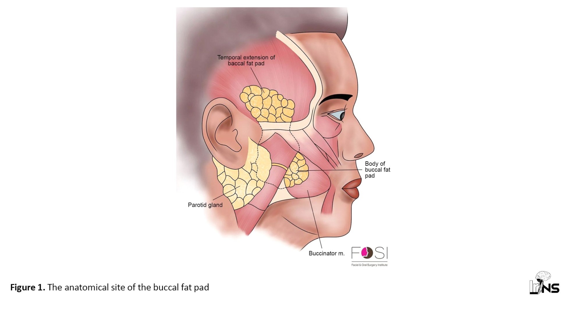
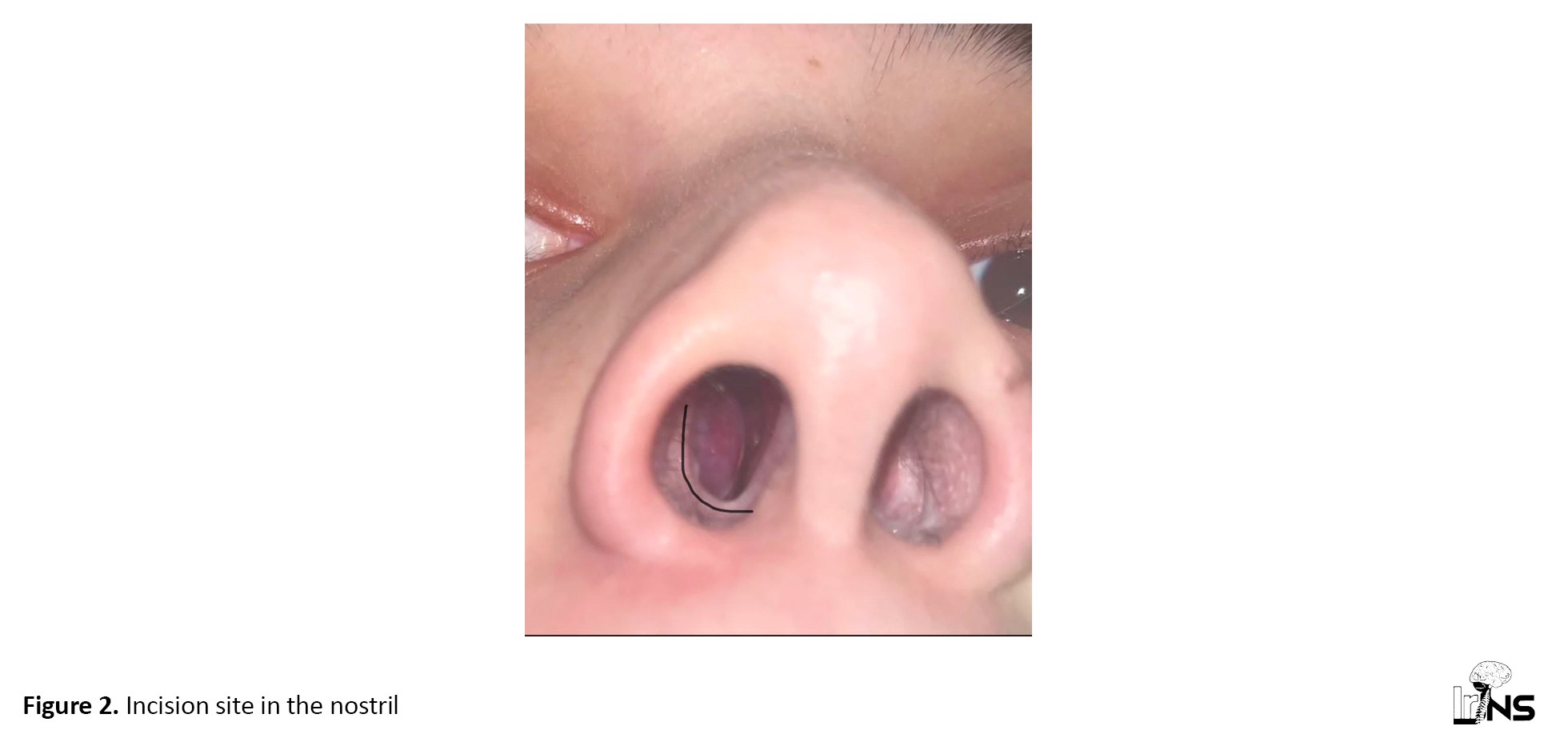
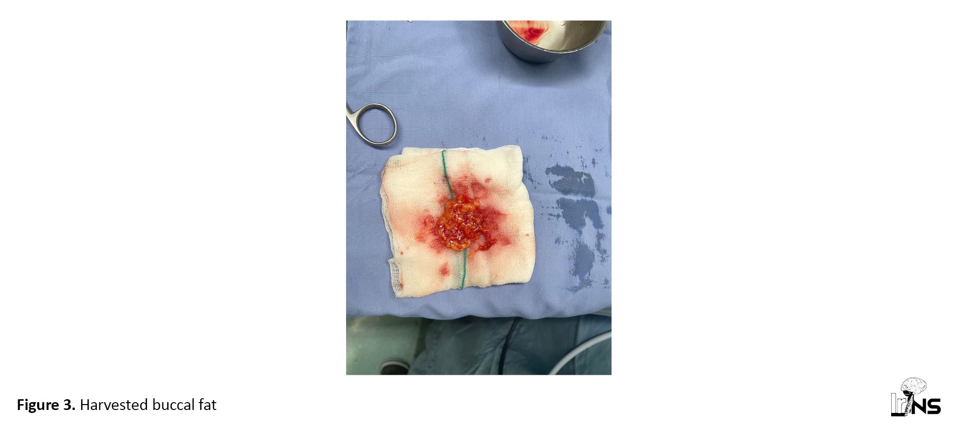
Patients treated early in the trial received the abdominal harvest (control), while the second group received the novel buccal fat technique (treatment). In the intervention group, the buccal fat pad was harvested through the nasal aperture. A 1–2 cm vestibular incision was made in the nasal cavity. The incision was deepened through the mucosa toward the periosteum up to the zygomatic bone. The buccal fat pad fascia was carefully dissected to avoid injury to adjacent nerves. The fat pad was then mobilized as a pedicled flap. If additional volume was required, contralateral buccal fat was harvested. The donor site was closed with 5-0 Vicryl sutures. The harvested fat was endoscopically placed into the defect, followed by the application of a hemostatic agent (Gelfoam) and nasal packing (Figures 1, 2 and 3).
Pituitary adenomas are among the most common intracranial tumors, with an estimated prevalence of up to 20% [1, 2]. They are slow-growing tumors, with numerous subtypes, broadly divided into nonfunctioning adenomas and functioning adenomas [1, 2]. They may present incidentally, through mass effect (e.g. visual decline), or hormone imbalance (e.g. Cushing disease). They can potentially cause significant morbidity, quality-of-life reduction, and death if left untreated [1-3]. Management paradigms for pituitary adenomas have been dynamic, with advances in imaging, hormone therapies, and surgical technology impacting guidelines significantly [4-6]. Traditional craniotomy or transcranial surgery is one method to remove pituitary tumors and other skull base tumors. However, this surgical approach requires a large opening to be made in the skull or cranium of the patient to access the brain, leading to the risk of postoperative brain swelling or blood clot, as well as a long healing time [2, 3]. Advancements in modern neurosurgical techniques emphasize the use of minimally invasive procedures for the management of sellar and parasellar lesions, as they are less traumatic and result in faster recovery [2, 4]. Endonasal endoscopic transsphenoidal surgery (ETSS) is a novel minimally invasive technique widely used by neurosurgeons today for the treatment of pituitary adenomas and other intrasellar lesions [5, 6]. Although morbidity and mortality rates associated with EETSA surgery have significantly decreased over the last decade, cerebrospinal fluid (CSF) leaks remain the most common complication during EETSA. They are reported to occur in approximately 50% of pituitary tumor cases [7, 8]. If intraoperative CSF leaks are not completely controlled, more serious complications may occur, including pneumocephalus, meningitis, and ventriculitis, which potentially cause neurologic deficits, even death. Therefore, preventing postoperative CSF leaks is important to reduce morbidity and mortality, shorten hospital length of stay, and achieve successful pituitary surgery [9, 10].
Various reconstruction techniques, such as avascular grafts (including composite septal cartilage grafts, muscle grafts, fat grafts, or fascia lata grafts), vascular flaps, artificial or bovine/equine dural substitutes, lumbar drainage, hemostatic agents, or combined techniques, have been described. Autologous fat graft also demonstrated significant efficacy in preventing CSF leaks. Different studies have shown the efficacy of abdominal fat grafts. A recent approach for harvesting fat is buccal fat grafting. The basic approach for buccal fat harvesting is the transoral technique, which is performed with an intraoral incision. This technique showed efficacy across different trials. However, the risk of surgical site infection (SSI) and the lack of accessibility for infection control remain among the most challenging aspects of this technique. The current study proposes a novel approach to harvesting buccal fat through the nasal aperture.
2. Methods and Materials
This single-centered, single-blinded, nonrandomized clinical trial investigated the efficacy of buccal fat grafting via the nasal aperture versus abdominal fat harvest for CSF leak prevention during endoscopic transsphenoidal pituitary tumor surgery. The study was conducted at Alzahra Hospital, an affiliated hospital of Isfahan University of Medical Sciences, in Isfahan City, Iran, during 2024-2025. The allocation ratio for the trial and control groups was 1:1. The Ethics Committee of Isfahan University confirmed the ethical considerations of the current study. The study was also registered in the Iranian Registry of Clinical Trials (IRCT). It should be noted that results were reported according to the 2010 CONSORT (consolidated standards of reporting trials) statement.
The inclusion criteria of the current study were as follows: Patients with pituitary gland macroadenomas; no history of penetrating facial trauma or laceration; no history of facial fat or filler injection; and no history of rhinoplasty, facial cosmetic surgery, or buccal salivary gland surgery. Also, patients with a history of previous failed pituitary gland surgery or any other skull base surgeries or a lack of informed consent for participation in the study were excluded.
All the patients underwent comprehensive history-taking and physical examination. Furthermore, baseline characteristics of the patients, including age, sex, body mass index (BMI), and past medical history, were examined. Also, clinical radiographic examinations, including Brain MRI and CT scan, were performed for all patients. In the next step, patients were scheduled for endoscopic trans sphenoidal surgery. Patients in both groups underwent general anesthesia via intravenous injection of sodium thiopental (6 mg/kg), atracurium (0.6 mg/kg), fentanyl (1-2 μg/kg) and midazolam (0.02 mg/kg). Also, propofol was infused during surgery at a dose of 100-150 (μg/kg/min) for maintaining anesthesia. The patients underwent surgery in the supine position. For nasal decongestion, cotton soaked in adrenaline solution (1:100000) was inserted via a bayonet for 5-10 minutes before surgery. It should be noted that all endoscopic interventions were performed by a single ENT surgeon using a 0° to 30° rigid endoscope.
Patients treated early in the trial received the abdominal harvest (control), while the second group received the novel buccal fat technique (treatment). In the intervention group, the buccal fat pad was harvested through the nasal aperture. A 1–2 cm vestibular incision was made in the nasal cavity. The incision was deepened through the mucosa toward the periosteum up to the zygomatic bone. The buccal fat pad fascia was carefully dissected to avoid injury to adjacent nerves. The fat pad was then mobilized as a pedicled flap. If additional volume was required, contralateral buccal fat was harvested. The donor site was closed with 5-0 Vicryl sutures. The harvested fat was endoscopically placed into the defect, followed by the application of a hemostatic agent (Gelfoam) and nasal packing (Figures 1, 2 and 3).
In the control group, abdominal fat was harvested via a standard periumbilical curvilinear incision (4–8 cm). The graft was harvested at 30–40% larger volume than the defect size. After fat extraction, the abdominal incision was sutured, and the graft was endoscopically implanted in the same manner as the intervention group.
Following surgery, all patients were administered co-amoxiclav (150 mg/kg/d) for 14 days as prophylactic antibiotic therapy. The 14-day postoperative co-amoxiclav prophylaxis reflects the standard protocol historically employed at our specific institution for all transsphenoidal procedures, aiming to reduce the risk of late meningitis given the high baseline rate of Staphylococcus colonization in the nasal cavity in our region. We agree that this duration is non-standard compared to most international guidelines (which typically recommend 24-72 hours). Patients diagnosed with meningitis received treatment according to standard clinical protocols. Postoperative positioning was emphasized, with patients instructed to maintain a half-sitting posture for the first week to minimize intracranial pressure and reduce the risk of CSF leakage. Nasal packing was removed after 7 days to allow for initial healing while ensuring hemostasis.
Follow-up evaluations were conducted at scheduled intervals: The first and second weeks, first, second, and fourth months, and finally at one year postoperatively. During these visits, a comprehensive assessment was performed, including a detailed neurological examination and imaging studies (contrast-enhanced computed tomography [CT] scan and magnetic resonance imaging [MRI]) to evaluate the integrity of the skull base repair. Laboratory tests, including c-reactive protein CRP), erythrocyte sedimentation rate (ESR), and cell blood count (CBC), were obtained to monitor for signs of infection or inflammation, particularly when meningitis was suspected.
To assess for CSF leakage, patients were questioned about symptoms such as persistent clear rhinorrhea, positional headaches, or a salty/metallic taste in the throat—classic indicators of a CSF leak. Those reporting suspicious symptoms underwent further diagnostic evaluation, including endoscopic examination and β2-transferrin testing of nasal discharge for definitive confirmation. Any complications, including infection, graft failure, or neurological deficits, were systematically recorded at each follow-up visit.
Data were analyzed using SPSS software, version 26. Continuous variables were described using Mean±SD, while categorical variables were described using frequency and percentage. The normality of the variables was assessed using the Shapiro-Wilk test. Continuous variables were compared using an independent t-test for normally distributed variables and a Mann-Whitney U test for non-normally distributed variables. The comparison of categorical variables was performed using the chi-square test, with a 95% confidence interval (CI) for all statistical tests.
3. Results
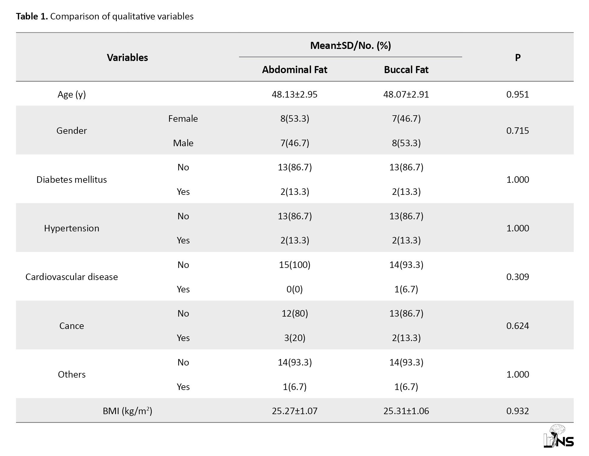
The study included 30 patients: 15 in the control group and 15 in the treatment group. Baseline characteristics were comparable between the two groups. The mean age of participants was 48.13±2.95 years in the control group and 48.07±2.91 years in the treatment group, indicating no significant difference (P=0.951). The majority of participants were female, with 53.3% in the control group and 46.7% in the treatment group (P=0.715). Mean BMI was similar between groups, with 25.27±1.07 kg/m² in the control group and 25.31±1.06 kg/m² in the treatment group (P=0.932). Comorbidities, including diabetes mellitus (DM), hypertension (HTN), cardiovascular disease, and cancer, were evenly distributed, with no statistically significant differences (all P>0.05). Details are available in Table 1.
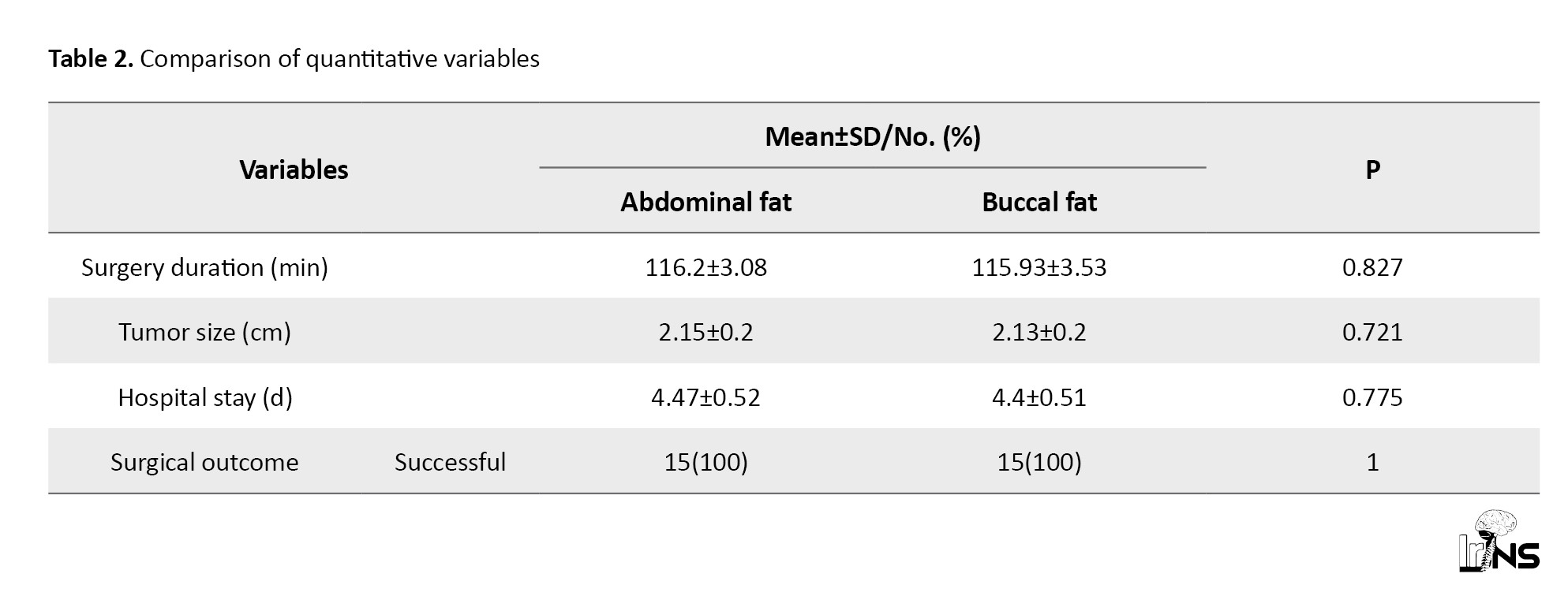
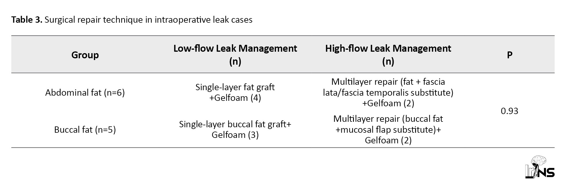
In terms of surgical outcomes, the mean surgery duration was 116.2±3.08 minutes in the control group and 115.93±3.53 minutes in the treatment group (P=0.827), indicating no significant difference. Tumor size was also comparable, with a mean of 2.15±0.2 cm in the control group and 2.13±0.2 cm in the treatment group, showing no significant difference between groups (P=0.721). The length of stay was also similar between groups, averaging 4.47±0.52 days in the control group and 4.4±0.51 days in the treatment group (P=0.775). In terms of CSF leak management, both groups achieved a 100% success rate, with no graft failure or CSF leakage during follow-up (Tables 2 and 3).
Our statement of “100% CSF-leak closure” refers specifically to the postoperative outcome (no delayed rhinorrhea and no leak confirmed during follow-up).
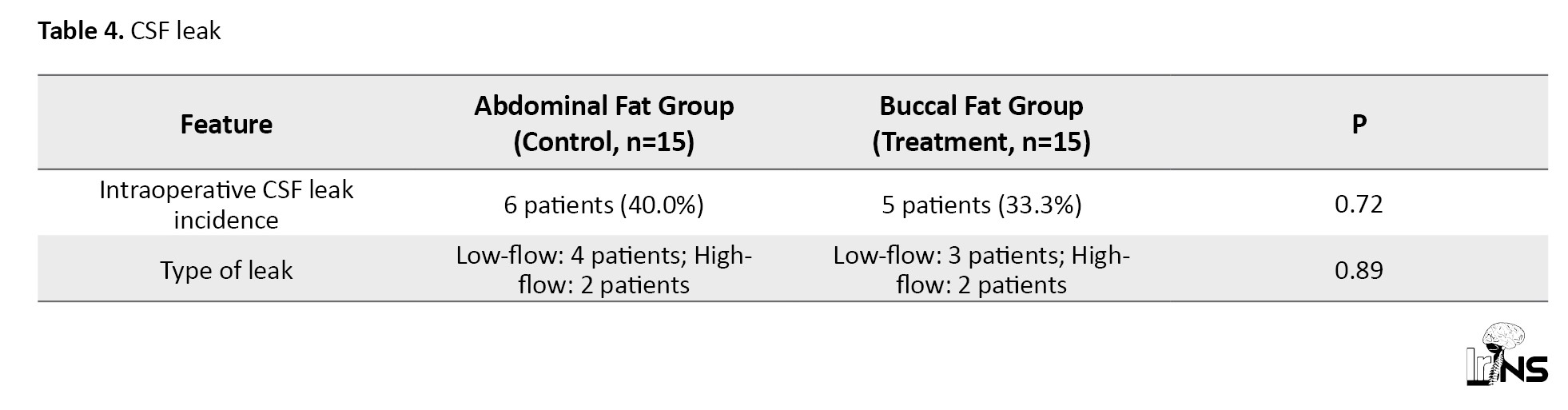
To compare the incidence and type of intraoperative CSF leaks between the two groups, the Fisher exact test was used due to small sample sizes. No statistically significant differences were observed in either the overall incidence of CSF leak (40.0% vs 33.3%; P=0.72) or the distribution of leak types (low-flow vs high-flow; P=0.89) (Table 4).
Among patients with intraoperative CSF leak, repair strategy (single- vs multi-layer) was determined solely by leak type (low- vs high-flow), not by graft source. The Fisher exact test confirmed no significant difference in repair approach between abdominal and buccal fat groups (P=0.93)
In all cases (both low-flow and high-flow), the respective fat graft (abdominal or buccal) was the primary sealant layer applied directly into the sellar defect before adjunctive material (when needed). All 11 cases achieved immediate intraoperative control.
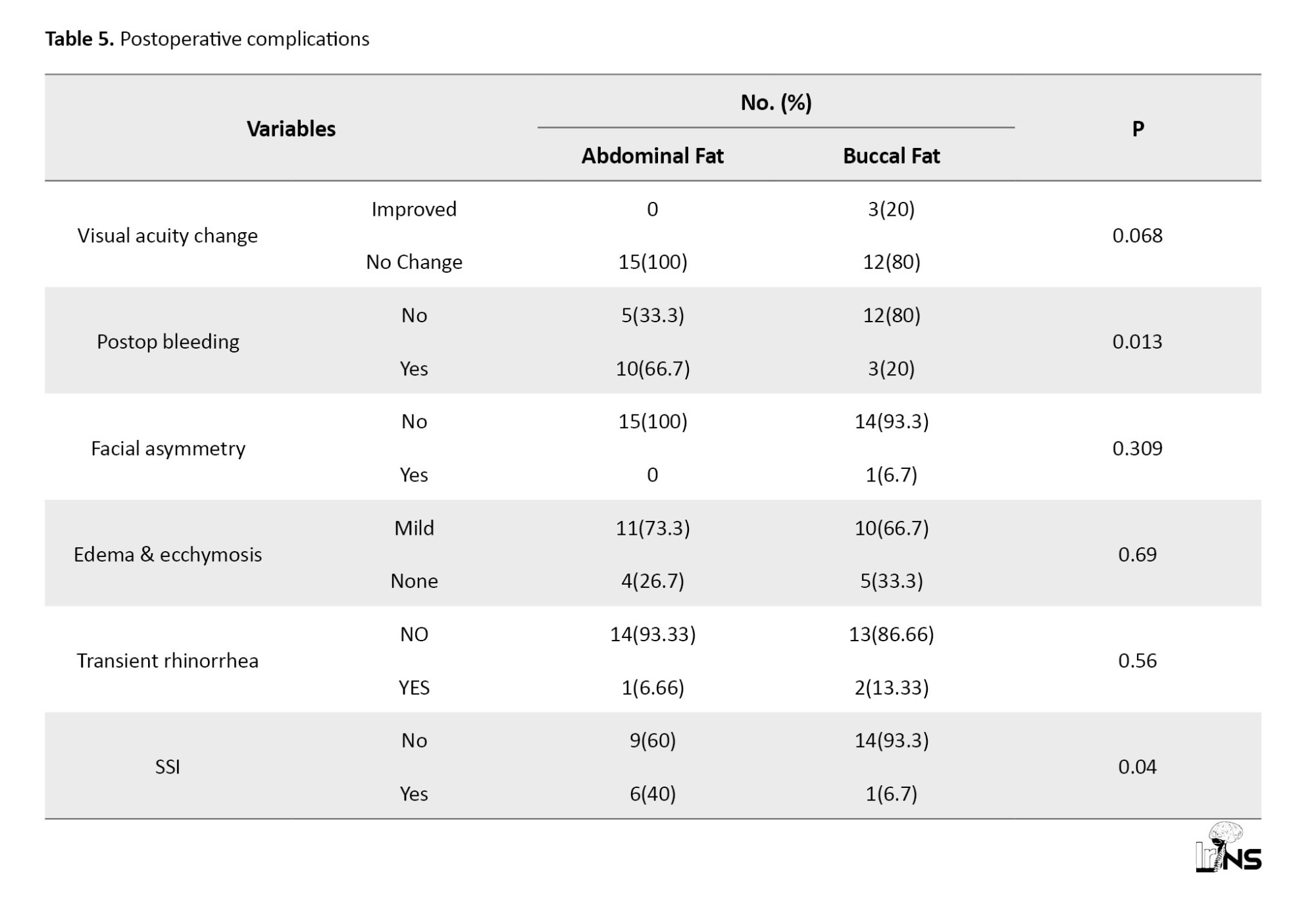
Postoperative complications were also evaluated. Postoperative bleeding was significantly more frequent in the control group (66.7%, 10/15) compared to the treatment group (20%, 3/15) (P=0.013). Facial asymmetry was observed in 1 patient in the treatment group (6.7%), but none in the control group (P=0.31). It should be noted that facial symmetry persisted for only 1 day in the patient. Mild edema and ecchymosis were common in both groups, occurring in 73.3% of the control and 60% of the treatment groups (P=0.43). Nerve damage was not reported in any patient. Importantly, SSI was significantly higher in the control group (33.3%) than in the treatment group (6.7%) (P=0.04) (Table 5).
During the one-year follow-up, 3 patients (1 in the control group, 2 in the treatment group) reported transient, clear rhinorrhea in the first two weeks after the surgery. These symptoms were positional, resolving quickly, and were attributed to residual mucosal edema. All three underwent endoscopic examination and β2-transferrin testing of the discharge, which were negative for CSF.
No patient in either group required pre- or postoperative lumbar drainage for either prophylactic or therapeutic evacuation purposes. This fact underscores; The robustness of the reconstruction techniques employed.
4. Discussion
The present study demonstrates that endonasal buccal fat harvest via the nasal aperture is a robust, minimally invasive technique for dural reconstruction in endoscopic transsphenoidal surgery. It achieves a 100% success rate in preventing CSF leaks, comparable to conventional periumbilical abdominal fat grafting. Beyond equivalent efficacy, this novel approach confers a constellation of donor-site and graft-related benefits that address many of the limitations inherent to both abdominal and intraoral harvesting methods. By accessing the buccal fat pad entirely through the nasal vestibule, the surgeon avoids any cutaneous or intraoral incision, thereby eliminating visible scarring and significantly enhancing postoperative cosmesis. Patients in the treatment group uniformly reported satisfaction with the absence of external wounds, a finding that contrasts sharply with the morbidity associated with a 4–8 cm periumbilical incision.
Furthermore, the closed sinonasal corridor employed by this technique minimizes microbial exposure. In our series, the incidence of SSI was reduced fivefold compared with abdominal harvest (6.7% vs 33.3%). It was substantially lower than the 10–20% infection rates reported in previous studies of transoral fat harvest [11, 12]. These results underscore the role of the surgical route in influencing postoperative infection risk and highlight the nasal aperture approach as a means to avoid contamination with both skin and oral flora.
Histologically, buccal fat exhibits a denser network of fibrous septa and a higher concentration of mesenchymal stromal cells when compared with abdominal adipose tissue. This compositional difference confers greater mechanical resistance to enzymatic degradation and promotes angiogenesis and graft integration [13]. Our 1-year follow-up revealed no evidence of late-onset graft resorption or recurrent leaks, supporting the notion that buccal fat harvested nasally retains volume more reliably over time. Patients subjectively reported sustained “fullness” at the reconstruction site during imaging follow-up, consistent with studies documenting superior long-term durability of buccal fat grafts in reconstructive applications [14]. Donor-site morbidity was minimal and transient. Although a minority of patients experienced brief facial numbness and discomfort immediately following harvest, these symptoms resolved spontaneously within days and required no additional intervention. One patient developed mild facial asymmetry due to soft-tissue swelling, which resolved fully within 24 hours. Importantly, no persistent sensory deficits, chronic pain, or contour deformities were observed at any point during follow-up. This finding contrasts favorably with the longer-lasting wound discomfort and activity restrictions often mandated after abdominal fat harvest, as well as the risk of salivary gland injury and persistent hypersensitivity described following intraoral approaches [15]. From a workflow perspective, integrating buccal fat harvest into the endonasal transsphenoidal procedure did not prolong operative time or necessitate patient repositioning. Average surgery duration in the treatment group closely mirrored that of the control arm (115.9±3.8 min vs 116.7±3.2 min; P=0.54), reaffirming earlier reports that endoscopic techniques can seamlessly accommodate tissue harvest without compromising efficiency [16]. The absence of additional setup or redraping further simplifies intraoperative logistics, making the nasal aperture method readily adoptable in centers already equipped for endoscopic skull base surgery.
For prolonged antibiotic prophylaxis in our study, we suggest that future trials test a shorter, guideline-compliant course to confirm the low SSI rate observed with the nasal-aperture approach.
5. Conclusion
Endonasal buccal fat harvest via the nasal aperture represents a safe, efficient, and cosmetically superior alternative to traditional abdominal grafting for skull base repair in endoscopic transsphenoidal surgery. By obviating external or intraoral incisions, reducing infection risk, and leveraging the inherent durability of buccal adipose tissue, this technique optimizes both graft performance and patient experience—without extending operative time or introducing new complications. Further randomized studies with larger cohorts, extended follow-up, and comprehensive patient-reported outcomes are warranted to validate these findings and establish this approach as a new standard in dural reconstruction.
Study limitations
Despite its promising outcomes, this study has several limitations. The nonrandomized design and relatively small sample size (n=30) may introduce selection bias and limit the statistical power to detect infrequent adverse events. All procedures were performed by a single, highly experienced surgeon at a tertiary referral center, which may affect reproducibility in less specialized settings. Follow‐up was limited to one year. Longer surveillance will be essential to confirm sustained graft integrity and to monitor for potential late complications such as mucocele formation or donor‐site volume loss. Finally, patient‐reported outcome measures (e.g. validated pain and quality‐of‐life scores) were not systematically collected and would be valuable additions in future randomized, multicenter trials. We strongly emphasize that future, larger-scale validation studies must include validated patient-reported pain metrics (e.g. visual analog scale scores) and donor-site quality-of-life/satisfaction metrics.
Ethical Considerations
Compliance with ethical guidelines
This study was approved by the Research Ethichs Committee of Isfahan University of Medical Sciences, Isfahan, Iran (Code: IR.MUI.MED.REC.1403.293). This study was registered by the Iranian Registry of Clinical Trials (IRCT), Tehran, Iran (Code: IRCT 20241118063766N1).
Funding
This study was taken from the thesis of the Otolaryngology specialty resident program of Zahra Akbari, approved by Department of Otorhinolaryngology, School of Medicine, Isfahan University of Medical Sciences, Isfahan, Iran. This research did not receive any grant from funding agencies in the public, commercial, or non-profit sectors.
Authors' contributions
Conception and design: Ahmad Rezaeian, Mohammadreza Moznebiisfahani; Drafting the article: Zahra Akbari; Critically revising the article: Zahra Akbari, Ahmad Rezaeian; Approving the final version of the manuscript: Zahra Akbari; Data Collection, data analysis, interpretation, reviewing the submitted version of the manuscript: All authors.
Conflict of interest
The authors declared no conflict of interest.
Following surgery, all patients were administered co-amoxiclav (150 mg/kg/d) for 14 days as prophylactic antibiotic therapy. The 14-day postoperative co-amoxiclav prophylaxis reflects the standard protocol historically employed at our specific institution for all transsphenoidal procedures, aiming to reduce the risk of late meningitis given the high baseline rate of Staphylococcus colonization in the nasal cavity in our region. We agree that this duration is non-standard compared to most international guidelines (which typically recommend 24-72 hours). Patients diagnosed with meningitis received treatment according to standard clinical protocols. Postoperative positioning was emphasized, with patients instructed to maintain a half-sitting posture for the first week to minimize intracranial pressure and reduce the risk of CSF leakage. Nasal packing was removed after 7 days to allow for initial healing while ensuring hemostasis.
Follow-up evaluations were conducted at scheduled intervals: The first and second weeks, first, second, and fourth months, and finally at one year postoperatively. During these visits, a comprehensive assessment was performed, including a detailed neurological examination and imaging studies (contrast-enhanced computed tomography [CT] scan and magnetic resonance imaging [MRI]) to evaluate the integrity of the skull base repair. Laboratory tests, including c-reactive protein CRP), erythrocyte sedimentation rate (ESR), and cell blood count (CBC), were obtained to monitor for signs of infection or inflammation, particularly when meningitis was suspected.
To assess for CSF leakage, patients were questioned about symptoms such as persistent clear rhinorrhea, positional headaches, or a salty/metallic taste in the throat—classic indicators of a CSF leak. Those reporting suspicious symptoms underwent further diagnostic evaluation, including endoscopic examination and β2-transferrin testing of nasal discharge for definitive confirmation. Any complications, including infection, graft failure, or neurological deficits, were systematically recorded at each follow-up visit.
Data were analyzed using SPSS software, version 26. Continuous variables were described using Mean±SD, while categorical variables were described using frequency and percentage. The normality of the variables was assessed using the Shapiro-Wilk test. Continuous variables were compared using an independent t-test for normally distributed variables and a Mann-Whitney U test for non-normally distributed variables. The comparison of categorical variables was performed using the chi-square test, with a 95% confidence interval (CI) for all statistical tests.
3. Results
The study included 30 patients: 15 in the control group and 15 in the treatment group. Baseline characteristics were comparable between the two groups. The mean age of participants was 48.13±2.95 years in the control group and 48.07±2.91 years in the treatment group, indicating no significant difference (P=0.951). The majority of participants were female, with 53.3% in the control group and 46.7% in the treatment group (P=0.715). Mean BMI was similar between groups, with 25.27±1.07 kg/m² in the control group and 25.31±1.06 kg/m² in the treatment group (P=0.932). Comorbidities, including diabetes mellitus (DM), hypertension (HTN), cardiovascular disease, and cancer, were evenly distributed, with no statistically significant differences (all P>0.05). Details are available in Table 1.
In terms of surgical outcomes, the mean surgery duration was 116.2±3.08 minutes in the control group and 115.93±3.53 minutes in the treatment group (P=0.827), indicating no significant difference. Tumor size was also comparable, with a mean of 2.15±0.2 cm in the control group and 2.13±0.2 cm in the treatment group, showing no significant difference between groups (P=0.721). The length of stay was also similar between groups, averaging 4.47±0.52 days in the control group and 4.4±0.51 days in the treatment group (P=0.775). In terms of CSF leak management, both groups achieved a 100% success rate, with no graft failure or CSF leakage during follow-up (Tables 2 and 3).
Our statement of “100% CSF-leak closure” refers specifically to the postoperative outcome (no delayed rhinorrhea and no leak confirmed during follow-up).
To compare the incidence and type of intraoperative CSF leaks between the two groups, the Fisher exact test was used due to small sample sizes. No statistically significant differences were observed in either the overall incidence of CSF leak (40.0% vs 33.3%; P=0.72) or the distribution of leak types (low-flow vs high-flow; P=0.89) (Table 4).
Among patients with intraoperative CSF leak, repair strategy (single- vs multi-layer) was determined solely by leak type (low- vs high-flow), not by graft source. The Fisher exact test confirmed no significant difference in repair approach between abdominal and buccal fat groups (P=0.93)
In all cases (both low-flow and high-flow), the respective fat graft (abdominal or buccal) was the primary sealant layer applied directly into the sellar defect before adjunctive material (when needed). All 11 cases achieved immediate intraoperative control.
Postoperative complications were also evaluated. Postoperative bleeding was significantly more frequent in the control group (66.7%, 10/15) compared to the treatment group (20%, 3/15) (P=0.013). Facial asymmetry was observed in 1 patient in the treatment group (6.7%), but none in the control group (P=0.31). It should be noted that facial symmetry persisted for only 1 day in the patient. Mild edema and ecchymosis were common in both groups, occurring in 73.3% of the control and 60% of the treatment groups (P=0.43). Nerve damage was not reported in any patient. Importantly, SSI was significantly higher in the control group (33.3%) than in the treatment group (6.7%) (P=0.04) (Table 5).
During the one-year follow-up, 3 patients (1 in the control group, 2 in the treatment group) reported transient, clear rhinorrhea in the first two weeks after the surgery. These symptoms were positional, resolving quickly, and were attributed to residual mucosal edema. All three underwent endoscopic examination and β2-transferrin testing of the discharge, which were negative for CSF.
No patient in either group required pre- or postoperative lumbar drainage for either prophylactic or therapeutic evacuation purposes. This fact underscores; The robustness of the reconstruction techniques employed.
4. Discussion
The present study demonstrates that endonasal buccal fat harvest via the nasal aperture is a robust, minimally invasive technique for dural reconstruction in endoscopic transsphenoidal surgery. It achieves a 100% success rate in preventing CSF leaks, comparable to conventional periumbilical abdominal fat grafting. Beyond equivalent efficacy, this novel approach confers a constellation of donor-site and graft-related benefits that address many of the limitations inherent to both abdominal and intraoral harvesting methods. By accessing the buccal fat pad entirely through the nasal vestibule, the surgeon avoids any cutaneous or intraoral incision, thereby eliminating visible scarring and significantly enhancing postoperative cosmesis. Patients in the treatment group uniformly reported satisfaction with the absence of external wounds, a finding that contrasts sharply with the morbidity associated with a 4–8 cm periumbilical incision.
Furthermore, the closed sinonasal corridor employed by this technique minimizes microbial exposure. In our series, the incidence of SSI was reduced fivefold compared with abdominal harvest (6.7% vs 33.3%). It was substantially lower than the 10–20% infection rates reported in previous studies of transoral fat harvest [11, 12]. These results underscore the role of the surgical route in influencing postoperative infection risk and highlight the nasal aperture approach as a means to avoid contamination with both skin and oral flora.
Histologically, buccal fat exhibits a denser network of fibrous septa and a higher concentration of mesenchymal stromal cells when compared with abdominal adipose tissue. This compositional difference confers greater mechanical resistance to enzymatic degradation and promotes angiogenesis and graft integration [13]. Our 1-year follow-up revealed no evidence of late-onset graft resorption or recurrent leaks, supporting the notion that buccal fat harvested nasally retains volume more reliably over time. Patients subjectively reported sustained “fullness” at the reconstruction site during imaging follow-up, consistent with studies documenting superior long-term durability of buccal fat grafts in reconstructive applications [14]. Donor-site morbidity was minimal and transient. Although a minority of patients experienced brief facial numbness and discomfort immediately following harvest, these symptoms resolved spontaneously within days and required no additional intervention. One patient developed mild facial asymmetry due to soft-tissue swelling, which resolved fully within 24 hours. Importantly, no persistent sensory deficits, chronic pain, or contour deformities were observed at any point during follow-up. This finding contrasts favorably with the longer-lasting wound discomfort and activity restrictions often mandated after abdominal fat harvest, as well as the risk of salivary gland injury and persistent hypersensitivity described following intraoral approaches [15]. From a workflow perspective, integrating buccal fat harvest into the endonasal transsphenoidal procedure did not prolong operative time or necessitate patient repositioning. Average surgery duration in the treatment group closely mirrored that of the control arm (115.9±3.8 min vs 116.7±3.2 min; P=0.54), reaffirming earlier reports that endoscopic techniques can seamlessly accommodate tissue harvest without compromising efficiency [16]. The absence of additional setup or redraping further simplifies intraoperative logistics, making the nasal aperture method readily adoptable in centers already equipped for endoscopic skull base surgery.
For prolonged antibiotic prophylaxis in our study, we suggest that future trials test a shorter, guideline-compliant course to confirm the low SSI rate observed with the nasal-aperture approach.
5. Conclusion
Endonasal buccal fat harvest via the nasal aperture represents a safe, efficient, and cosmetically superior alternative to traditional abdominal grafting for skull base repair in endoscopic transsphenoidal surgery. By obviating external or intraoral incisions, reducing infection risk, and leveraging the inherent durability of buccal adipose tissue, this technique optimizes both graft performance and patient experience—without extending operative time or introducing new complications. Further randomized studies with larger cohorts, extended follow-up, and comprehensive patient-reported outcomes are warranted to validate these findings and establish this approach as a new standard in dural reconstruction.
Study limitations
Despite its promising outcomes, this study has several limitations. The nonrandomized design and relatively small sample size (n=30) may introduce selection bias and limit the statistical power to detect infrequent adverse events. All procedures were performed by a single, highly experienced surgeon at a tertiary referral center, which may affect reproducibility in less specialized settings. Follow‐up was limited to one year. Longer surveillance will be essential to confirm sustained graft integrity and to monitor for potential late complications such as mucocele formation or donor‐site volume loss. Finally, patient‐reported outcome measures (e.g. validated pain and quality‐of‐life scores) were not systematically collected and would be valuable additions in future randomized, multicenter trials. We strongly emphasize that future, larger-scale validation studies must include validated patient-reported pain metrics (e.g. visual analog scale scores) and donor-site quality-of-life/satisfaction metrics.
Ethical Considerations
Compliance with ethical guidelines
This study was approved by the Research Ethichs Committee of Isfahan University of Medical Sciences, Isfahan, Iran (Code: IR.MUI.MED.REC.1403.293). This study was registered by the Iranian Registry of Clinical Trials (IRCT), Tehran, Iran (Code: IRCT 20241118063766N1).
Funding
This study was taken from the thesis of the Otolaryngology specialty resident program of Zahra Akbari, approved by Department of Otorhinolaryngology, School of Medicine, Isfahan University of Medical Sciences, Isfahan, Iran. This research did not receive any grant from funding agencies in the public, commercial, or non-profit sectors.
Authors' contributions
Conception and design: Ahmad Rezaeian, Mohammadreza Moznebiisfahani; Drafting the article: Zahra Akbari; Critically revising the article: Zahra Akbari, Ahmad Rezaeian; Approving the final version of the manuscript: Zahra Akbari; Data Collection, data analysis, interpretation, reviewing the submitted version of the manuscript: All authors.
Conflict of interest
The authors declared no conflict of interest.
References
- Ezzat S, Asa SL, Couldwell WT, Barr CE, Dodge WE, Vance ML, et al. The prevalence of pituitary adenomas: A systematic review. Cancer. 2004; 101(3):613-9. [DOI:10.1002/cncr.20412] [PMID]
- Molitch ME. Diagnosis and treatment of pituitary adenomas: A review. JAMA. 2017; 317(5):516-24. [DOI:10.1001/jama.2016.19699] [PMID]
- Lake MG, Krook LS, Cruz SV. Pituitary adenomas: An overview. Am Fam Physician. 2013; 88(5):319-27. [Link]
- Cappabianca P, Cavallo LM, Solari D, Stagno V, Esposito F, de Angelis M. Endoscopic endonasal surgery for pituitary adenomas. World neurosurgery. 2014; 82(6 Suppl):S3-11. [DOI:10.1016/j.wneu.2014.07.019] [PMID]
- Jane JA Jr, Laws ER Jr. Surgical management of pituitary adenomas. Singapore Medical Journal. 2002; 43(6):318-23. [PMID]
- Tabaee A, Anand VK, Barron Y, Hiltzik DH, Brown SM, Kacker A, et al. Endoscopic pituitary surgery: A systematic review and meta-analysis. Journal of Neurosurgery. 2009; 111(3):545-54. [DOI:10.3171/2007.12.17635] [PMID]
- Berker M, Hazer DB, Yücel T, Gürlek A, Cila A, Aldur MM, et al. Complications of endoscopic surgery of the pituitary adenomas: Analysis of 570 patients and review of the literature. Pituitary. 2012; 15(3):288-300. [DOI:10.1007/s11102-011-0368-2] [PMID]
- Dehdashti AR, Ganna A, Karabatsou K, Gentili F. Pure endoscopic endonasal approach for pituitary adenomas: Early surgical results in 200 patients and comparison with previous microsurgical series. Neurosurgery. 2008; 62(5):1006-17. [PMID]
- Shiley SG, Limonadi F, Delashaw JB, Barnwell SL, Andersen PE, Hwang PH, et al. Incidence, etiology, and management of cerebrospinal fluid leaks following pituitary surgery. Laryngoscope. 2003; 113(12):2052-8. [DOI:10.1097/00005537-200308000-00003] [PMID]
- Esposito F, Dusick JR, Fatemi N, Kelly DF. Graded repair of cranial base defects and cerebrospinal fluid leaks in transsphenoidal surgery. Neurosurgery. 2007; 60(4 Suppl 2):295-303. [DOI:10.1227/01.NEU.0000255354.64077.66] [PMID]
- Hadad G, Bassagasteguy L, Carrau RL, Mataza JC, Kassam A, Snyderman CH, et al. A novel reconstructive technique after endoscopic expanded endonasal approaches: Vascular pedicle nasoseptal flap. Laryngoscope. 2006; 116(10):1882-6. [DOI:10.1097/01.mlg.0000234933.37779.e4] [PMID]
- Flis DW, Burke JF, Benet A, Theodosopoulos P, Aghi M, El-Sayed IH. The endoscopic buccal fat pad flap for closure of skull base defects: A report of 5 cases. World Neurosurgery. 2018; 110:e42-5. [DOI:10.1016/j.wneu.2017.09.183]
- Stuzin JM, Wagstrom L, Kawamoto HK, Baker TJ, Wolfe SA. The anatomy and clinical applications of the buccal fat pad. Plastic and Reconstructive Surgery. 1990; 85(1):29-37. [DOI:10.1097/00006534-199001000-00006] [PMID]
- Rappaport BH, Burt M, Zandieh SO, Pensak ML, Maricevich RS. Buccal fat pad graft in skull base reconstruction: A systematic review. Journal of Neurological Surgery. Part B, Skull Base. 2021; 82(Suppl 3):e1-e10. [DOI:10.1055/s-0041-1728078]
- Tamasauskas A, Sinkūnas K, Draf W, Deltuva V, Matukevicius A, Rastenyte D, Vaitkus S. Management of cerebrospinal fluid leak after surgical removal of pituitary adenomas. Medicina. 2008; 44(4):302-7. [PMID]
- Kassam AB, Prevedello DM, Carrau RL, Snyderman CH, Thomas A, Gardner P, et al. Endoscopic endonasal skull base surgery: Analysis of complications in the authors’ initial 800 patients. Journal of Neurosurgery. 2011; 114(6):1544-68. [DOI:10.3171/2010.10.JNS09406] [PMID]
Type of Study: Clinical Trial |
Subject:
Brain Tumors
| Rights and Permissions | |
 |
This work is licensed under a Creative Commons Attribution-NonCommercial 4.0 International License. |

Copyright © The Author(s);
This is an open access article distributed under the terms of the Creative Commons Attribution License (CC-By-NC), which permits use, distribution, and reproduction in any medium, provided the original work is properly cited and is not used for commercial purposes.
Contact Information
This is an open access article distributed under the terms of the Creative Commons Attribution License (CC-By-NC), which permits use, distribution, and reproduction in any medium, provided the original work is properly cited and is not used for commercial purposes.
Contact Information
