Sun, Nov 22, 2026
Volume 12 - Contnious Publishing
Iran J Neurosurg 2026, 12 - Contnious Publishing: 0-0 |
Back to browse issues page



Download citation:
BibTeX | RIS | EndNote | Medlars | ProCite | Reference Manager | RefWorks
Send citation to:


BibTeX | RIS | EndNote | Medlars | ProCite | Reference Manager | RefWorks
Send citation to:
Baradaran-Bagheri A, Mohazzab-Torabi S. Posterior Only Approach for T1–T2 disc herniation, Case Report and Literature Review. , Iran J Neurosurg. 2026; 12 : 4
URL: http://irjns.org/article-1-536-en.html
URL: http://irjns.org/article-1-536-en.html


1- Department of Neurosurgery, Faculty of Medicine, Alborz University of Medical Sciences, Karaj, Iran.
2- Department of Neurosurgery, Faculty of Medicine, Alborz University of Medical Sciences, Karaj, Iran. ,smntrb@gmail.com
2- Department of Neurosurgery, Faculty of Medicine, Alborz University of Medical Sciences, Karaj, Iran. ,
Full Text [PDF 1990 kb]
(211 Downloads)
| Abstract (HTML) (619 Views)
Full Text: (44 Views)
1. Background and Importance
Thoracic disc herniations are rare clinical entities, accounting for approximately 0.25-0.75% of all intervertebral disc ruptures [1]. The majority occur in the lower thoracic spine, most commonly at T11–T12, whereas lesions at the upper thoracic levels (T1–T4) are exceedingly uncommon [2]. Among these, T1–T2 disc herniations comprise only 1-3% of thoracic disc herniations, and fewer than 40 cases have been reported in the literature since the first description in 1945.
The clinical spectrum of T1–T2 varies with the degree and direction of neural compression. Central or anterolateral herniations may produce spinal cord compression and myelopathy, manifesting as paraparesis, gait disturbance, or sphincter dysfunction [3]. Conversely, posterolateral lesions often impinge on the exiting T1 nerve root, leading to radiculopathy characterized by pain radiating to the medial arm, axilla, and hand, sometimes associated with Horner’s syndrome due to involvement of the sympathetic chain [4]. Because of the narrow thoracic canal, even a relatively small disc extrusion at this level can produce disproportionately severe neurological deficits, underscoring the importance of early recognition and intervention. From a surgical perspective, the upper thoracic spine presents unique anatomical and biomechanical challenges. The region is difficult to access via an anterior approach due to the proximity of the sternum and mediastinal structures, while posterior approaches may risk destabilization if not performed with pedicle-sparing techniques [5, 6]. Moreover, the cervicothoracic junction is a transitional zone subject to altered biomechanics, especially in patients with prior cervical fusion, which may predispose to adjacent segment degeneration at C7–T1 and T1–T2 [7]. Consequently, the choice between anterior, posterior, or motion-preserving approaches must be individualized, balancing the extent of neural compression, anatomical accessibility, and the long-term impact on spinal stability.
2. Case Presentation
A 37-year-old woman presented with progressive cervicothoracic pain and right upper-limb weakness that had failed to improve with conservative management. She reported increasing difficulty with hand function, particularly medial arm and shoulder pain, and paresthesias with radiculopathy that significantly interfered with daily activities. Neurological examination demonstrated weakness of the intrinsic muscles of the right hand, sensory loss in the T1 dermatome, and asymmetrical hyperreflexia. No signs of myelopathy such as gait disturbance or sphincter involvement were detected.
Magnetic resonance imaging revealed a large centrolateral disc herniation at the T1–T2 level, associated with marked foraminal narrowing and compression of the exiting nerve root (Figure 1).
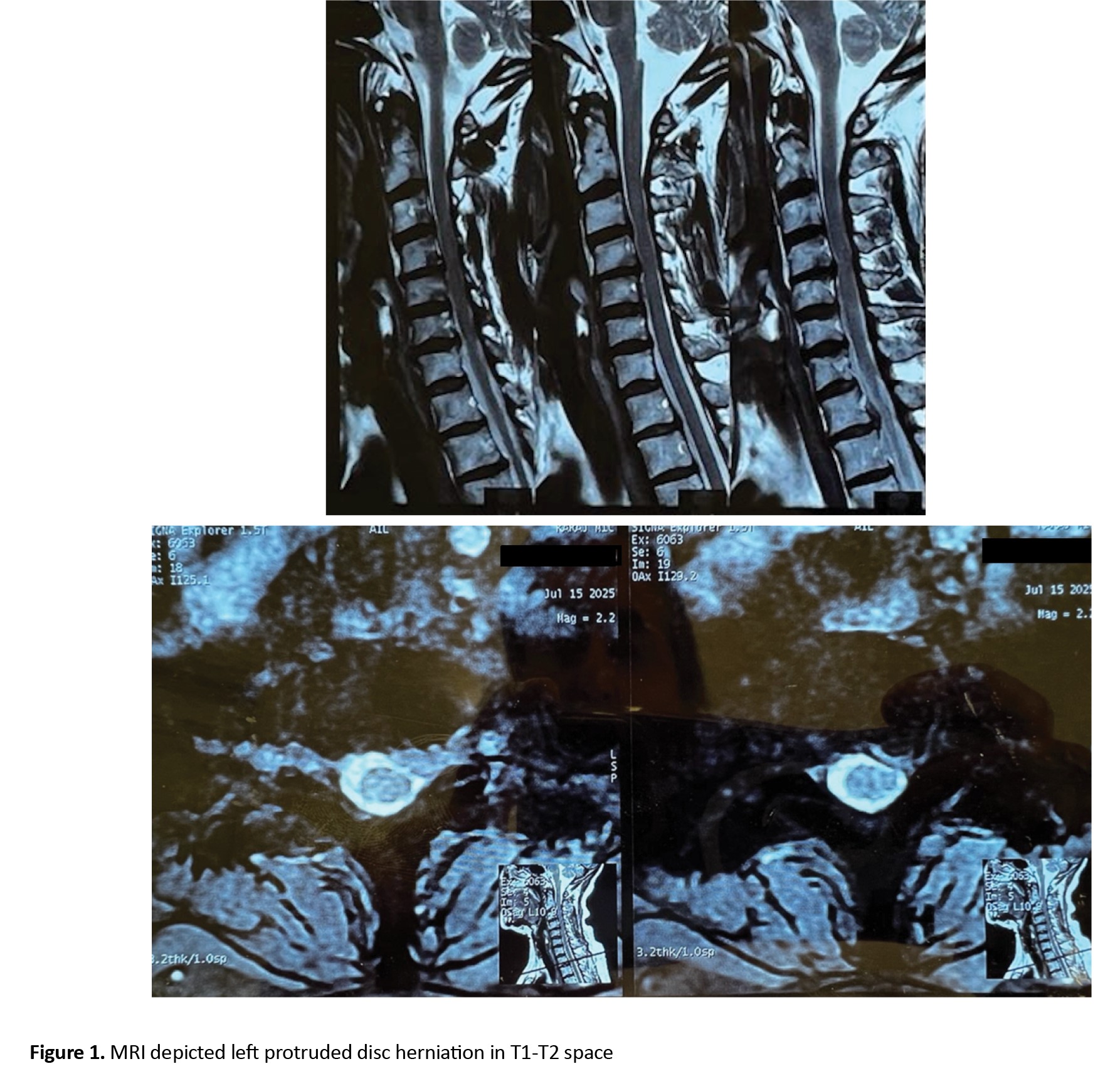 Additional imaging also demonstrated mild degenerative changes at the adjacent C7–T1 disc space, but without significant cord compression. Given the persistence of symptoms, the progression of neurological deficits, and the lack of response to non-operative therapy, the decision was made to proceed with surgical intervention.
After weighing considerations, we selected a posterior motion-preserving approach that allowed full neural decompression without adding unnecessary biomechanical stress. The operative plan consisted of a T1 laminectomy combined with bilateral foraminotomies and microsurgical excision of the extruded disc, performed under continuous intraoperative neuromonitoring to minimize the risk of cord or root injury.
The patient was placed under general anesthesia and positioned prone on a radiolucent frame with careful padding of dependent areas. Somatosensory and motor evoked potentials were monitored throughout the procedure. A midline incision was made extending across the cervicothoracic junction, followed by subperiosteal exposure of the posterior elements from C7 to T2.
A T1 laminectomy was performed using high-speed drilling and Kerrison rongeurs to gain access to the spinal canal. Subsequently, unilateral foraminotomies at T1–T2 were performed to enlarge the neural foramina. Under the operating microscope and using meticulous microsurgical technique, the fragment was mobilized and removed en bloc. Adequacy of decompression was confirmed by clearance of the foraminal pathway. Hemostasis was secured with bipolar cautery, and hemostatic material was applied over a drain. Throughout the operation, neuromonitoring signals remained stable, and no complications occurred. Recovery was uneventful. The patient demonstrated immediate improvement in grip strength and relief of radicular pain. Sensory changes in the T1 dermatome progressively improved over subsequent weeks. At follow-up, she remained neurologically intact, with no new deficits or signs of myelopathy. Postoperative magnetic resonance imaging (MRI) confirmed complete removal of the disc fragment and satisfactory decompression of both the cord and exiting nerve root.
3. Discussion
Symptomatic intervertebral disc herniation at the T1–T2 level is exceptionally rare, accounting for only a small fraction of thoracic disc herniations [8]. The cervicothoracic junction represents a complex anatomical transition between the mobile cervical and rigid thoracic spine, characterized by unique biomechanical and neurovascular challenges [9]. This region’s deep location, narrow operative corridor, and proximity to critical mediastinal structures make surgical management both technically demanding and conceptually nuanced [10].
In the present case, multiple surgical options were carefully analyzed. Purely anterior approaches—while providing the most direct access to the ventral pathology—pose significant technical difficulties due to the limited working corridor and the risk of injury to adjacent mediastinal structures, including the trachea, esophagus, and great vessels [11, 12]. Moreover, anterior fusion across T1–T2 may lead to biomechanical isolation of the cervicothoracic junction, predisposing the adjacent C7–T1 motion segment to accelerated degenerative change due to altered stress distribution [13].
Conversely, limited posterior approaches such as keyhole foraminotomy, though minimally invasive, are insufficient for safe and complete resection of large paracentral or central disc fragments [14]. Extensive posterior fusion involving both C7–T1 and T1–T2 levels would likely impose unnecessary morbidity, including reduced cervical mobility and adjacent segment disease [15]. Hence, the selected approach must ensure adequate ventral decompression while preserving the stability and kinematics of the transitional segment [16].
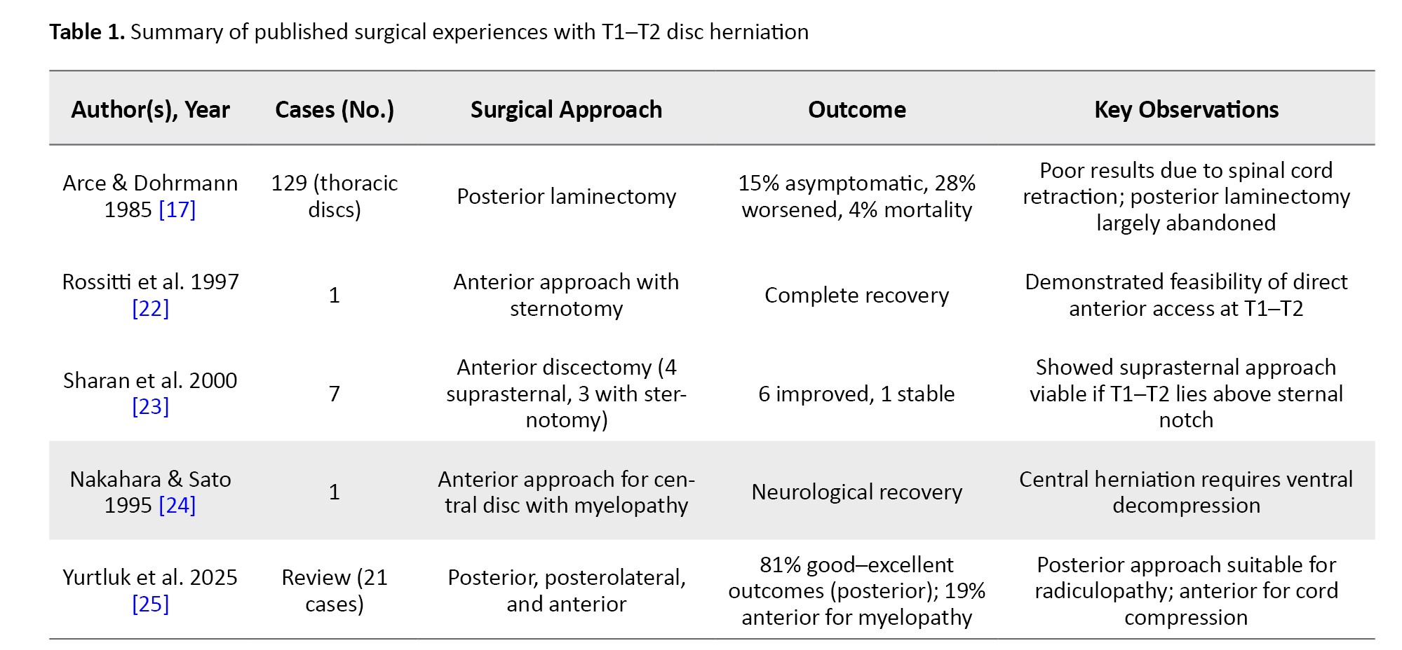
Historically, posterior decompressive laminectomy was the primary method for thoracic disc herniation; however, the outcomes were poor. In the seminal series by Arce and Dohrmann involving 129 patients, only 15% were asymptomatic postoperatively, while 28% worsened and 4% died. These suboptimal results were attributed to excessive spinal cord manipulation required for disc removal through a posterior trajectory [17]. Improved outcomes—over 80% of patients were asymptomatic or improved—were achieved in those treated via lateral, posterolateral, or transthoracic routes, leading to the near abandonment of posterior laminectomy as a sole decompressive technique for ventrally located thoracic discs.
T1–T2 disc herniation often presents with clinical features that mimic cervical radiculopathy or thoracic outlet syndrome, contributing to frequent diagnostic delays. The predominant symptoms include neck, shoulder, and arm pain (approximately 60%), sensory disturbances (23%), and motor weakness (18%). Radiculopathy is the most common presentation (up to 87% of reported cases), while Horner’s syndrome—secondary to sympathetic chain disruption—occurs in approximately 21–24% of patients [15, 16, 18]. In Table 1, we demonstrated multiple case series about T1 disc herniations.
Anatomically, the T1 root contributes to the medial cord of the brachial plexus, forming components of the ulnar and median nerves and the medial cutaneous nerve of the forearm [19]. T1 radiculopathy manifests as paresthesia along the medial forearm and weakness of intrinsic hand muscles—often mimicking C8 radiculopathy or distal ulnar neuropathies. Preservation of deep finger flexion with isolated intrinsic weakness suggests T1 involvement, a critical diagnostic clue distinguishing it from C8 pathology [20].
The T1–T2 level is often obscured radiographically by the shoulder girdle and may be excluded from conventional cervical MRI studies. Consequently, in patients presenting with lower cervical radiculopathy accompanied by atypical features or Horner’s syndrome, MRI should be extended to include the upper thoracic segments [21]. The differential diagnosis is broad, encompassing Pancoast tumors, apical lung masses, and sympathetic chain or intramedullary lesions. Comprehensive imaging, therefore, plays a pivotal role not only in diagnosis but also in surgical planning by delineating the relationship between the lesion and the sternal notch, a determinant of anterior approach feasibility.
These data collectively suggest that:
1) Posterior laminectomy alone yields inferior outcomes due to inadequate ventral decompression and cord manipulation. 2) Anterior or anterolateral approaches are superior for central or calcified herniations causing myelopathy. 3) Posterior or posterolateral routes (transfacet, transpedicular, or lateral extracavitary) remain appropriate for paracentral or foraminal herniations causing radiculopathy. 4) Minimally invasive and endoscopic techniques increasingly offer effective decompression with reduced morbidity, though their application at T1–T2 remains limited to specialized centers.
In the present case, MRI revealed a paracentral, sequestered disc fragment displacing the spinal cord laterally and compressing the T1 root. The coexistence of radiculopathy and Horner’s syndrome suggested preganglionic sympathetic dysfunction secondary to root compression at the intermediolateral cell column. Therefore, an posterier discectomy was chosen to achieve total decompression of both the T1 root and the sympathetic fibers traversing this region. The immediate postoperative resolution of Horner’s syndrome corroborates the effectiveness of direct decompression and supports the theory that complete removal of ventral pathology is essential for restoring sympathetic function.
4. Conclusion
T1–T2 intervertebral disc herniation remains a rare but clinically important differential diagnosis in patients presenting with lower cervical radiculopathy or unexplained Horner’s syndrome. Due to the anatomical constraints of the cervicothoracic junction, meticulous preoperative imaging and individualized surgical planning are crucial. While posterior approaches may suffice for lateralized lesions producing radiculopathy, anterior or anterolateral approaches provide superior access for central herniations causing cord compression or autonomic dysfunction.
Ultimately, optimal outcomes depend on achieving total neural decompression while minimizing biomechanical compromise of the transitional junction. Early recognition and an anatomically precise, pathology-specific approach remain the cornerstones of management for this uncommon yet surgically challenging condition.
Ethical Considerations
Compliance with ethical guidelines
Written informed consent was obtained from the patient for publication of this case report and accompanying images. The patient was informed that all identifying information would be removed to protect anonymity.
Funding
This research did not receive any specific grants from funding agencies in the public, commercial, or not-for-profit sectors.
Authors' contributions
Conceptualization and study design: Ali Baradaran-Bagheri; Data collection and writing the original draft: Saman Mohazzab-Torabi; Data analysis, interpretation, review, editing, and final approval: All authors.
Conflict of interest
The authors declared no conflict of interest.
References
Thoracic disc herniations are rare clinical entities, accounting for approximately 0.25-0.75% of all intervertebral disc ruptures [1]. The majority occur in the lower thoracic spine, most commonly at T11–T12, whereas lesions at the upper thoracic levels (T1–T4) are exceedingly uncommon [2]. Among these, T1–T2 disc herniations comprise only 1-3% of thoracic disc herniations, and fewer than 40 cases have been reported in the literature since the first description in 1945.
The clinical spectrum of T1–T2 varies with the degree and direction of neural compression. Central or anterolateral herniations may produce spinal cord compression and myelopathy, manifesting as paraparesis, gait disturbance, or sphincter dysfunction [3]. Conversely, posterolateral lesions often impinge on the exiting T1 nerve root, leading to radiculopathy characterized by pain radiating to the medial arm, axilla, and hand, sometimes associated with Horner’s syndrome due to involvement of the sympathetic chain [4]. Because of the narrow thoracic canal, even a relatively small disc extrusion at this level can produce disproportionately severe neurological deficits, underscoring the importance of early recognition and intervention. From a surgical perspective, the upper thoracic spine presents unique anatomical and biomechanical challenges. The region is difficult to access via an anterior approach due to the proximity of the sternum and mediastinal structures, while posterior approaches may risk destabilization if not performed with pedicle-sparing techniques [5, 6]. Moreover, the cervicothoracic junction is a transitional zone subject to altered biomechanics, especially in patients with prior cervical fusion, which may predispose to adjacent segment degeneration at C7–T1 and T1–T2 [7]. Consequently, the choice between anterior, posterior, or motion-preserving approaches must be individualized, balancing the extent of neural compression, anatomical accessibility, and the long-term impact on spinal stability.
2. Case Presentation
A 37-year-old woman presented with progressive cervicothoracic pain and right upper-limb weakness that had failed to improve with conservative management. She reported increasing difficulty with hand function, particularly medial arm and shoulder pain, and paresthesias with radiculopathy that significantly interfered with daily activities. Neurological examination demonstrated weakness of the intrinsic muscles of the right hand, sensory loss in the T1 dermatome, and asymmetrical hyperreflexia. No signs of myelopathy such as gait disturbance or sphincter involvement were detected.
Magnetic resonance imaging revealed a large centrolateral disc herniation at the T1–T2 level, associated with marked foraminal narrowing and compression of the exiting nerve root (Figure 1).
After weighing considerations, we selected a posterior motion-preserving approach that allowed full neural decompression without adding unnecessary biomechanical stress. The operative plan consisted of a T1 laminectomy combined with bilateral foraminotomies and microsurgical excision of the extruded disc, performed under continuous intraoperative neuromonitoring to minimize the risk of cord or root injury.
The patient was placed under general anesthesia and positioned prone on a radiolucent frame with careful padding of dependent areas. Somatosensory and motor evoked potentials were monitored throughout the procedure. A midline incision was made extending across the cervicothoracic junction, followed by subperiosteal exposure of the posterior elements from C7 to T2.
A T1 laminectomy was performed using high-speed drilling and Kerrison rongeurs to gain access to the spinal canal. Subsequently, unilateral foraminotomies at T1–T2 were performed to enlarge the neural foramina. Under the operating microscope and using meticulous microsurgical technique, the fragment was mobilized and removed en bloc. Adequacy of decompression was confirmed by clearance of the foraminal pathway. Hemostasis was secured with bipolar cautery, and hemostatic material was applied over a drain. Throughout the operation, neuromonitoring signals remained stable, and no complications occurred. Recovery was uneventful. The patient demonstrated immediate improvement in grip strength and relief of radicular pain. Sensory changes in the T1 dermatome progressively improved over subsequent weeks. At follow-up, she remained neurologically intact, with no new deficits or signs of myelopathy. Postoperative magnetic resonance imaging (MRI) confirmed complete removal of the disc fragment and satisfactory decompression of both the cord and exiting nerve root.
3. Discussion
Symptomatic intervertebral disc herniation at the T1–T2 level is exceptionally rare, accounting for only a small fraction of thoracic disc herniations [8]. The cervicothoracic junction represents a complex anatomical transition between the mobile cervical and rigid thoracic spine, characterized by unique biomechanical and neurovascular challenges [9]. This region’s deep location, narrow operative corridor, and proximity to critical mediastinal structures make surgical management both technically demanding and conceptually nuanced [10].
In the present case, multiple surgical options were carefully analyzed. Purely anterior approaches—while providing the most direct access to the ventral pathology—pose significant technical difficulties due to the limited working corridor and the risk of injury to adjacent mediastinal structures, including the trachea, esophagus, and great vessels [11, 12]. Moreover, anterior fusion across T1–T2 may lead to biomechanical isolation of the cervicothoracic junction, predisposing the adjacent C7–T1 motion segment to accelerated degenerative change due to altered stress distribution [13].
Conversely, limited posterior approaches such as keyhole foraminotomy, though minimally invasive, are insufficient for safe and complete resection of large paracentral or central disc fragments [14]. Extensive posterior fusion involving both C7–T1 and T1–T2 levels would likely impose unnecessary morbidity, including reduced cervical mobility and adjacent segment disease [15]. Hence, the selected approach must ensure adequate ventral decompression while preserving the stability and kinematics of the transitional segment [16].
Historically, posterior decompressive laminectomy was the primary method for thoracic disc herniation; however, the outcomes were poor. In the seminal series by Arce and Dohrmann involving 129 patients, only 15% were asymptomatic postoperatively, while 28% worsened and 4% died. These suboptimal results were attributed to excessive spinal cord manipulation required for disc removal through a posterior trajectory [17]. Improved outcomes—over 80% of patients were asymptomatic or improved—were achieved in those treated via lateral, posterolateral, or transthoracic routes, leading to the near abandonment of posterior laminectomy as a sole decompressive technique for ventrally located thoracic discs.
T1–T2 disc herniation often presents with clinical features that mimic cervical radiculopathy or thoracic outlet syndrome, contributing to frequent diagnostic delays. The predominant symptoms include neck, shoulder, and arm pain (approximately 60%), sensory disturbances (23%), and motor weakness (18%). Radiculopathy is the most common presentation (up to 87% of reported cases), while Horner’s syndrome—secondary to sympathetic chain disruption—occurs in approximately 21–24% of patients [15, 16, 18]. In Table 1, we demonstrated multiple case series about T1 disc herniations.
Anatomically, the T1 root contributes to the medial cord of the brachial plexus, forming components of the ulnar and median nerves and the medial cutaneous nerve of the forearm [19]. T1 radiculopathy manifests as paresthesia along the medial forearm and weakness of intrinsic hand muscles—often mimicking C8 radiculopathy or distal ulnar neuropathies. Preservation of deep finger flexion with isolated intrinsic weakness suggests T1 involvement, a critical diagnostic clue distinguishing it from C8 pathology [20].
The T1–T2 level is often obscured radiographically by the shoulder girdle and may be excluded from conventional cervical MRI studies. Consequently, in patients presenting with lower cervical radiculopathy accompanied by atypical features or Horner’s syndrome, MRI should be extended to include the upper thoracic segments [21]. The differential diagnosis is broad, encompassing Pancoast tumors, apical lung masses, and sympathetic chain or intramedullary lesions. Comprehensive imaging, therefore, plays a pivotal role not only in diagnosis but also in surgical planning by delineating the relationship between the lesion and the sternal notch, a determinant of anterior approach feasibility.
These data collectively suggest that:
1) Posterior laminectomy alone yields inferior outcomes due to inadequate ventral decompression and cord manipulation. 2) Anterior or anterolateral approaches are superior for central or calcified herniations causing myelopathy. 3) Posterior or posterolateral routes (transfacet, transpedicular, or lateral extracavitary) remain appropriate for paracentral or foraminal herniations causing radiculopathy. 4) Minimally invasive and endoscopic techniques increasingly offer effective decompression with reduced morbidity, though their application at T1–T2 remains limited to specialized centers.
In the present case, MRI revealed a paracentral, sequestered disc fragment displacing the spinal cord laterally and compressing the T1 root. The coexistence of radiculopathy and Horner’s syndrome suggested preganglionic sympathetic dysfunction secondary to root compression at the intermediolateral cell column. Therefore, an posterier discectomy was chosen to achieve total decompression of both the T1 root and the sympathetic fibers traversing this region. The immediate postoperative resolution of Horner’s syndrome corroborates the effectiveness of direct decompression and supports the theory that complete removal of ventral pathology is essential for restoring sympathetic function.
4. Conclusion
T1–T2 intervertebral disc herniation remains a rare but clinically important differential diagnosis in patients presenting with lower cervical radiculopathy or unexplained Horner’s syndrome. Due to the anatomical constraints of the cervicothoracic junction, meticulous preoperative imaging and individualized surgical planning are crucial. While posterior approaches may suffice for lateralized lesions producing radiculopathy, anterior or anterolateral approaches provide superior access for central herniations causing cord compression or autonomic dysfunction.
Ultimately, optimal outcomes depend on achieving total neural decompression while minimizing biomechanical compromise of the transitional junction. Early recognition and an anatomically precise, pathology-specific approach remain the cornerstones of management for this uncommon yet surgically challenging condition.
Ethical Considerations
Compliance with ethical guidelines
Written informed consent was obtained from the patient for publication of this case report and accompanying images. The patient was informed that all identifying information would be removed to protect anonymity.
Funding
This research did not receive any specific grants from funding agencies in the public, commercial, or not-for-profit sectors.
Authors' contributions
Conceptualization and study design: Ali Baradaran-Bagheri; Data collection and writing the original draft: Saman Mohazzab-Torabi; Data analysis, interpretation, review, editing, and final approval: All authors.
Conflict of interest
The authors declared no conflict of interest.
References
- Cornips E. Thoracic disc herniations: an underestimated disease in search for a surgical cure [doctoral thesis]. Antwerp: University of Antwerp; 2023. [Link]
- Lafage R, Steinberger J, Pesenti S, Assi A, Elysee JC, Iyer S, et al. Understanding thoracic spine morphology, shape, and proportionality. Spine. 2020; 45(3):149-57. [DOI:10.1097/BRS.0000000000003227] [PMID]
- Jacobson R, Osen A. Spinal cord disease. Hankey’s clinical neurology. Boca Raton: CRC Press; 2020. [DOI:10.1201/9780429299476-23]
- Radecki J, Feinberg JH, Zimmer ZR. T1 radiculopathy: electrodiagnostic evaluation. Hss Journal. 2009;5(1):73-7. [DOI:10.1007/s11420-008-9105-4] [PMID] [PMCID]
- Kawashima M, Rhoton Jr AL, Matsushima T. Comparison of posterior approaches to the posterior incisural space: microsurgical anatomy and proposal of a new method, the occipital bi-transtentorial/falcine approach. Neurosurgery. 2008; 62(6):SHC1136-SHC49. [DOI:10.1227/01.NEU.0000333780.12058.44] [PMID]
- Pointillart V, Aurouer N, Gangnet N, Vital J-M. Anterior approach to the cervicothoracic junction without sternotomy: A report of 37 cases. Spine. 2007; 32(25):2875-9. [DOI:10.1097/BRS.0b013e31815b7632] [PMID]
- McDonald CL, Badida R, Alsoof D, Daher M, Homer A, Crisco JJ, et al. Crossing the cervicothoracic junction: a biomechanical investigation of C7: Versus: T1 caudal selection’s effect on adjacent segment motion in posterior cervical fusion. Spine. 2024; 49(24):1743-9. [DOI:10.1097/BRS.0000000000005129] [PMID]
- Vanichkachorn JS, Vaccaro AR. Thoracic disk disease: diagnosis and treatment. JAAOS-Journal of the American Academy of Orthopaedic Surgeons. 2000; 8(3):159-69. [DOI:10.5435/00124635-200005000-00003] [PMID]
- Negovetiæ L, Èerina V, Sajko T, Glaviæ Z. Intradural disc herniation at the T1-T2 level. Croatian Medical Journal. 2001; 42(193):5. [Link]
- Morgan H, Abood C. Disc herniation at T1-2: Report of four cases and literature review. Journal of neurosurgery. 1998; 88(1):148-50. [DOI:10.3171/jns.1998.88.1.0148] [PMID]
- Bouthors C, Benzakour A, Court C. Surgical treatment of thoracic disc herniation: An overview. International Orthopaedics. 2019; 43(4):807-16. [DOI:10.1007/s00264-018-4224-0] [PMID]
- Son ES, Lee SH, Park SY, Kim KT, Kang CH, Cho SW. Surgical treatment of t1-2 disc herniation with t1 radiculopathy: A case report with review of the literature. Asian Spine Journal. 2012; 6(3):199. [DOI:10.4184/asj.2012.6.3.199] [PMID] [PMCID]
- Anto M, Manuel A, Jayachandran A, Thomas SG, Joseph A, Thankachan A, et al. Horner’s syndrome secondary to T1-T2 intervertebral disc prolapse. Surgical Neurology International. 2022; 13:412. [DOI:10.25259/SNI_580_2022] [PMID] [PMCID]
- Foss-Skiftesvik J, Hougaard MG, Larsen VA, Hansen K. Clinical Reasoning: Partial Horner syndrome and upper right limb symptoms following chiropractic manipulation. Neurology. 2015; 84(21):e175-80. [DOI:10.1212/WNL.0000000000001616] [PMID]
- Possley D, Luczak SB, Angus A, Montgomery D. T1-T2 disk herniation presenting with Horner syndrome: A case report with literary review. JAAOS Global Research & Reviews. 2018; 2(11):e016. [DOI:10.5435/JAAOSGlobal-D-18-00016] [PMID] [PMCID]
- Teixeira JC, Santos MM, Melancia JL. T1-T2 Herniated disk presenting with Horner syndrome. World Neurosurgery. 2017; 107:1050. [DOI:10.1016/j.wneu.2017.08.069] [PMID]
- Arce CA, Dohrmann GJ. Thoracic disc herniation: improved diagnosis with computed tomographic scanning and a review of the literature. Surgical Neurology. 1985; 23(4):356-61. [DOI:10.1016/0090-3019(85)90206-X] [PMID]
- Gokcen HB, Erdogan S, Gumussuyu G, Ozturk S, Ozturk C. A rare case of T1-2 thoracic disc herniation mimicking cervical radiculopathy. International Journal of Spine Surgery. 2017; 11(4):30. [DOI:10.14444/4030] [PMID] [PMCID]
- Funayama T, Noguchi H, Abe T, Miura K, Nagashima K, Kumagai H, et al. A case of thoracic disc herniation characterized by marked posture-related dynamic changes in neurological symptoms. Interdisciplinary Neurosurgery. 2018; 14:1-4. [DOI:10.1016/j.inat.2018.05.015]
- Randhawa PS, Roark C, Case D, Seinfeld J. Idiopathic spinal cord herniation associated with a thoracic disc herniation: Case report, surgical video, and literature review. Clinical Spine Surgery. 2020; 33(6):222-9. [DOI:10.1097/BSD.0000000000000896] [PMID] [PMCID]
- Ozturk C, Tezer M, Sirvanci M, Sarier M, Aydogan M, Hamzaoglu A. Far lateral thoracic disc herniation presenting with flank pain. The Spine Journal. 2006; 6(2):201-3. [DOI:10.1016/j.spinee.2005.08.004] [PMID]
- Rossitti S, Stephensen H, Ekholm S, von Essen C. The anterior approach to high thoracic (T1-T2) disc herniation. British Journal of Neurosurgery. 1993; 7(2):189-92. [DOI:10.3109/02688699309103477] [PMID]
- Sharan AD, Przybylski GJ, Tartaglino L. Approaching the upper thoracic vertebrae without sternotomy or thoracotomy: A radiographic analysis with clinical application. Spine. 2000; 25(8):910-16. [DOI:10.1097/00007632-200004150-00003] [PMID]
- Nakahara S, Sato T. First thoracic disc herniation with myelopathy. European Spine Journal. 1995; 4(6):366-367. [DOI:10.1007/BF00300300] [PMID]
- Yurtluk MD, APAYDIN AS, Kina H, Than K. Thoracic disc herniations: diagnosis, surgical techniques, and complication insights. Turkish Journal of Medical Sciences. 2025; 55(1):17-23. [DOI:10.55730/1300-0144.5939] [PMID] [PMCID]
Type of Study: Case report |
Subject:
Spine
Send email to the article author
| Rights and Permissions | |
 |
This work is licensed under a Creative Commons Attribution-NonCommercial 4.0 International License. |

Copyright © The Author(s);
This is an open access article distributed under the terms of the Creative Commons Attribution License (CC-By-NC), which permits use, distribution, and reproduction in any medium, provided the original work is properly cited and is not used for commercial purposes.
Contact Information
This is an open access article distributed under the terms of the Creative Commons Attribution License (CC-By-NC), which permits use, distribution, and reproduction in any medium, provided the original work is properly cited and is not used for commercial purposes.
Contact Information
